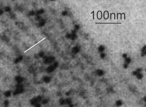
This research study uses what they call surfactant A filamentous structure and lipid bilayers to study whether surfactant protein A is responsible for some of the functional and structural features of surfactant. In one of their figures I tried to match their banding pattern with that found in vivo in the various animals I have looked at searching for an identity for this RER granule (or intracisternal body) and the numbers don’t match up completely. Their banding in vitro between surfactant protein A filaments and lipid bilayer material is more than the 100 nm periodicity I find in vivo in guinea pig and ferret and dog alveolar type II cells. I am certainly not challenging their measurements, I would more likely challenge my own, since I have not photographed a grid pattern in decades with which to calibrate the TEM. SO, that said, I conclude that there is the possibility that something in surfactant A is at work here, and the observation which is much more interesting is that they describe intersections at right angles and bran ching in vitro… just like the branching and curving that is found in the granules (intracisternal bodies) in vivo.
Nades Palaniyar, Ross A. Ridsdale, Stephen A. Hearn, Yew Meng Heng, F. Peter Ottensmeyer, Fred Possmayer, George Harauz American Journal of Physiology – Lung Cellular and Molecular Physiology Published 1 April 1999 Vol. 276 no. 4, L631-L641
This is a crop and edit of one of their pix. The white 100 nm bar is mine, made from their scale marker of 50 nm.