Three different organizations of this protein, some parallel some perpendicular to the plane of the nuclear envelope.

Three different organizations of this protein, some parallel some perpendicular to the plane of the nuclear envelope.
I continued to be interested in this image of a guinea pig alveolar type Ii cell nucleus, just off to the side which had a couple areas that looked for all the world as if they were part of some coiling of (coiling of the 30 nm coil) and maybe even linked together in a similar fasion as the nucleosomes themselves, but maybe packages of 3 or 4 nucleosomes with a linking region that looked to be consistent as well. Video is primitive, but it does give relevancy to the big-beads (to be distinguished from the more commonly used “beads” on a string for nucleosomally wound DNA. I really have scoured the internet and science articles to find another who has seen this kind of arrangement of chromatin at the periphery of the nucleus…. I have not found much. Nucleosomes are typically NOT as large as these one-step-further- wound DNA. Video is here:
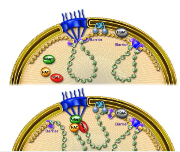
Here is an awesome picture (diagram) which i found after making my short video…. the link to the diagram and those who made it is here. www.cell.com/molecular-cell/fulltext/S1097-2765%2810%2900172-3
The intracisternal body shows up in the RER (the membrane of the RER is the dark boundary of the central profile and there is also some bounded by the outer nuclear membrane) but still not in sufficient detail to be absolutely sure how many bands are between the major divisions. I have highlighted these in cyan, maybe you can figure out how many are there, I think it might be 7 including both sides of the major bands (of which there are 5 in this profile of RER.
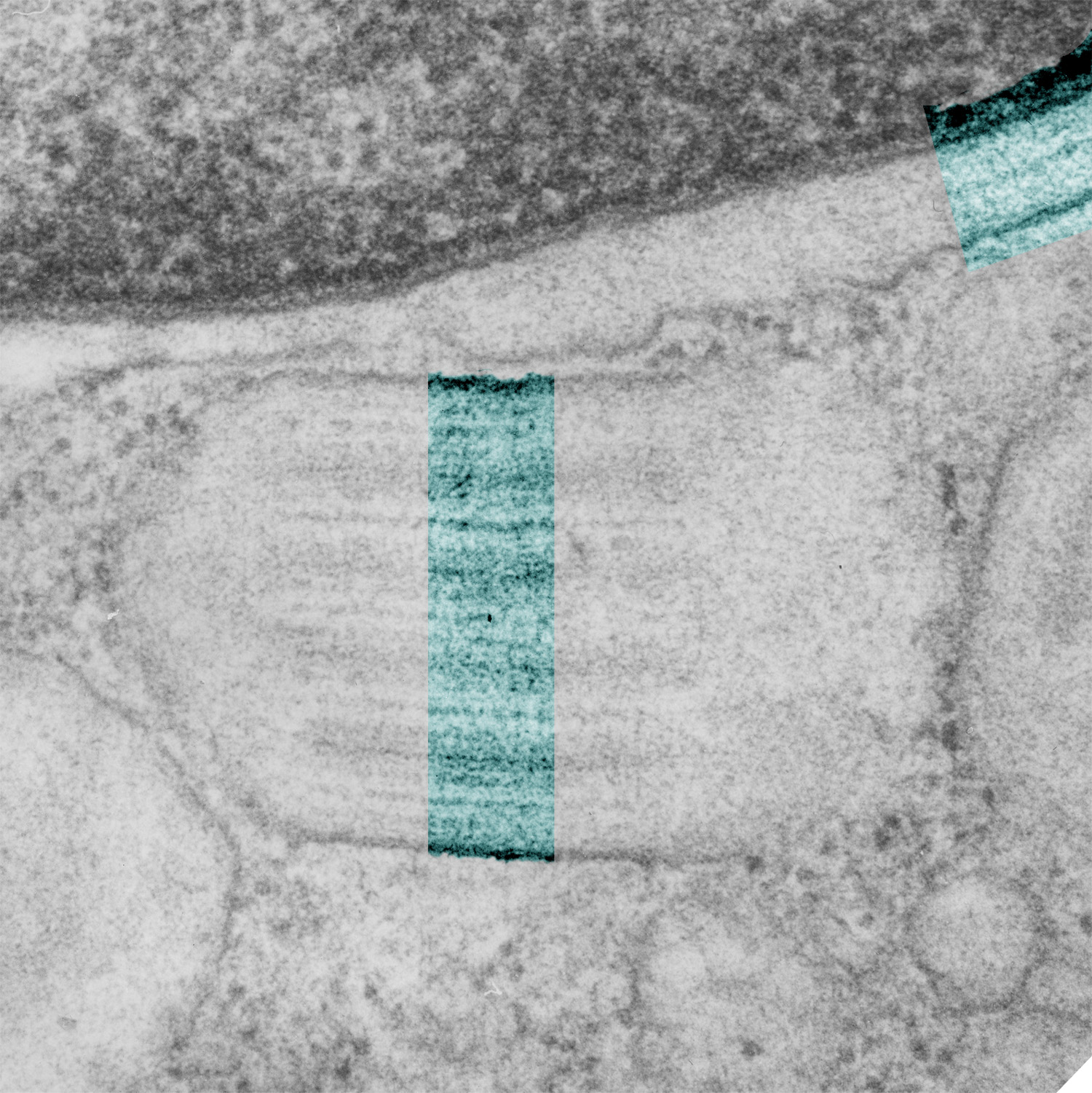
I have added additional images and a pretty high magnification view of an intracisternal protein body in the type II alveolar cell of a guinea pig (Animal 301, aged, Hartley) which was used as a control in some exposure studies. I think it seems pretty clear (that is a cya statement) that there are two ribosomes positioned just inside the dominant bands in these highly structured protein inclusions (see the last part of this video on youTube). The dominant bands are spaced just about 0.1 microns apart. HERE is the link to youtube.
The journal article where the cisternal body is discussed in detail is an “oldie” published in the Journal of Ultrastructure and Molecular Structure Research, 95: 131-141, 1986.
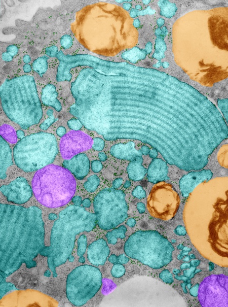
Working to determine whether there is an order to the ribosomes at the growing ends of the intracisternal proteins that are found in some aged animals of this and other species, I pseudocolored another transmission electron micrograph, intracisternal protein bodies are purple, and like the other images, the ribosomesa re cyan, and the lamellar bodies in the type II pneumocytes are gold-orange colored. It seems like there might be about two ribosomes on either side of the scalloped growin end, just before the darkest of the bands running perpendicular to the growing end. I can provide links to images that are very high resolution (26 megs) if you ask.
I am still trying to figure out what the ratio of ribosomes on the elongating end of these RER structures is. I think that the ribosomes appear to be staggered over the odd number of inner bands. Here is a jpg of a type II cell from a guinea pig (aged) which actually has a huge number of separate RER profiles stuffed with this protein (which I have pseudocolored cyan) and a few mitochondrial profiles (pseudocolored purple) a couple of lamellar bodies (kind of a gold color) and again, ribosomes here and there and plentiful at that, between and at the elongating ends of the profiles of RER.
The paracrystal inclusion in the ferret (and guinea pig and mongrel dog) rough endoplasmic reticulum of the type II alveolar pneumocyte is really a fun structure. I have been examining old transmission electron micrographs in an effort to uncover just a little more about its structure and occurrence. I don’t think I realized back in 1986 when i published a peer review manuscript on this structure that it could be found in cross section to look like a tube, with a central point and concentric rings (see post several days ago). So looking for more confirmation on that aspect of the body I decided to make this short animated gif which shows the original electron micrograph and fades into a pseudocolored and enhanced electron micrograph where the ribosomes that feed the intracisternal protein body are highlighted in cyan and the intersection of two perpendicular alignments I have used the “burn” tool in photoshop to enhance. NO structures were deleted, this is just processing of the original micrograph. 
For several decades I have been wanting to study these intracisternal protein bodies, since they are spectacularly organized, seem to grow from two ends, and have large ribosome-protein complexes at the growing end and can sometimes be seen in cross section as concentric rings with a central point. I pseudo-colored this ferret type II cell in with colors. It is from a collection of photomicrographs which depict these bodies and an especially interesting and organized sample. High resolution images are available on two separate image libraries, but here is a thumbnail image.
