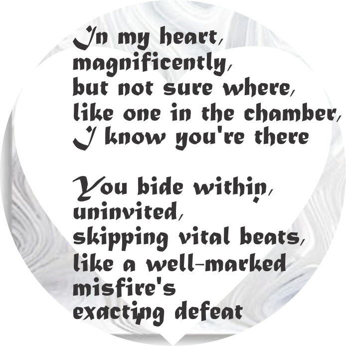
In my heart,
magnificiently,
but not sure where,
like one in the chamber,
I know your’re there
You abide within,
uninvited,
skipping vital beats,
like a well-marked
misfire’s
exacting defeat.
sara louise miller witt, 301602016
In my heart,
magnificiently,
but not sure where,
like one in the chamber,
I know your’re there
You abide within,
uninvited,
skipping vital beats,
like a well-marked
misfire’s
exacting defeat.
sara louise miller witt, 301602016
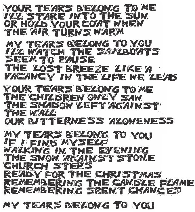 Your tears belong to me
i’ll stare into the sun
or hold your coat when
the air turns warm
My tears belong to you
i’ll watch the sailboats
seem to pause
the lost breeze
like a vacancy
in the life we lead
Your tears belong to me
the children only saw
the shadow left against
the wall
our bitterness aloneness
my tears belong to you
if i find myself walking
in the evening
the snow against
stone church steps
ready for the christmas
remembering the candle flame
remembering spent chances
words, RLB, stamps, mm
 words, RLB, hand cut stamps, mm
Type II alveolar pneumocyte here pseudocolored as follows: nucleus, purple; nuclear pores light green; perichromatin granules, violet-red; intracisternal bodies, even in the perinuclear space, light blue (these are looking more and more like they might be over production of Surfactant protein A – a collectin which has modest surfactant properties in the alveolar space but probably greater impact on pulmonary immune function. Interestingly it lines up pretty much on the button with two mirror sets of the actual surfactant protein A molecule (which is a group of 18 similar single protein units spanning about 50 nm, so two top to bottom would be about 100 nm the width of one set of bands in an intracisternal body — at least I hope it works out that way); lamellar bodies, of which there are four, gold; and a single mitochondrion, green.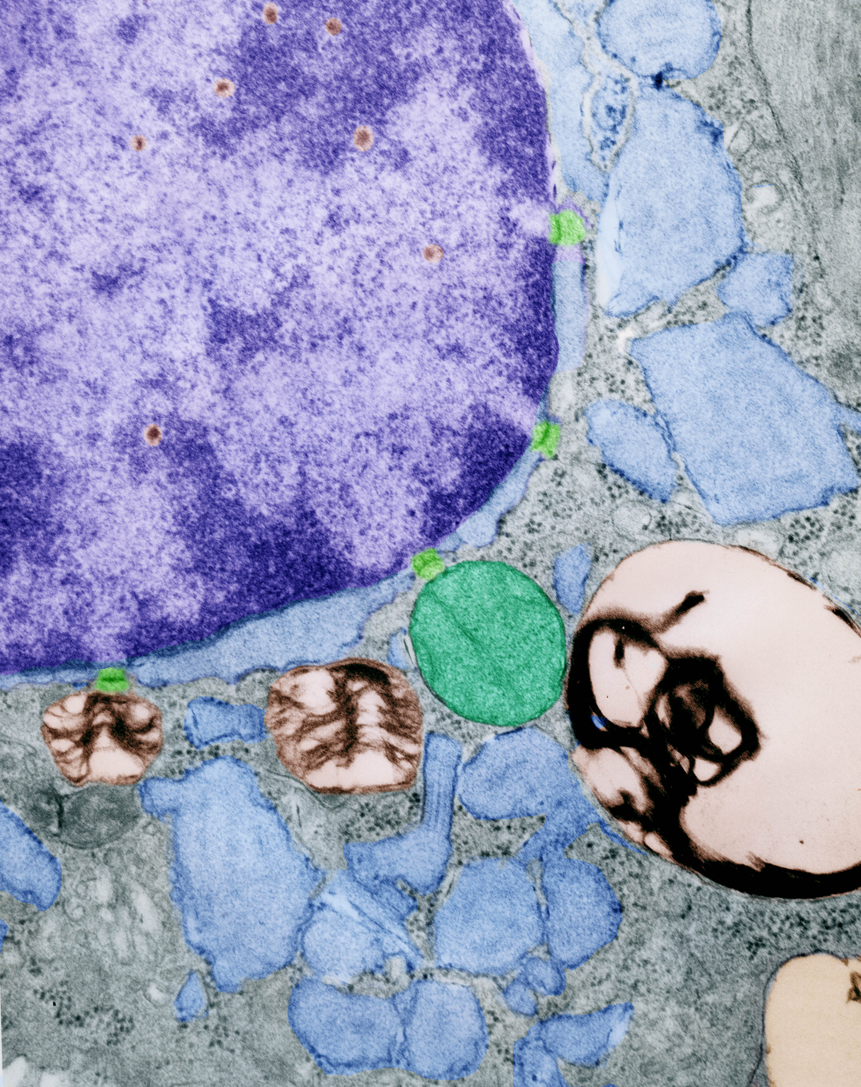
Still probing the literature to see whether I can match up a protein to the paracrystalline pattern seen in electron micrographs of guinea pig, ferret and sometimes in dog lung, I found a paper which might shed some light on the issue. “
If you kissed me really
For the first time.
It would be the only
Reason for being in a limo.
If you kissed me madly
It would give me purpose
For riding in a taxi.
If you threw your
Arm around me, it
Would be the only
Rein that stopped me,
with top rolled back,
From being lifted to the sky.
If I remembered you,
On this bus ride home,
I’d shiver in the sun,
knowing that the ride was
About to end.
RLB Midnight 11/21/2011
PDF, midnight_11_21_2011
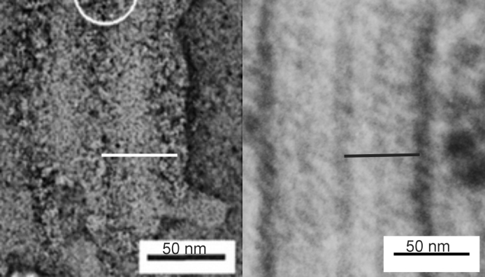
Here is a short video of the intracisternal protein organization that I have found in guinea pig (and a couple other mammals) in the alveolar type II pneumocytes using transmission electron microscopy. It seems that in the most advantageous photographs, and with the method of preservation that was used on these tissues (glutaraldehyde – paraformaldehyde fix, with osmium tetroxide post fix, either Epon 812 or Spurr’s resin and lead and uranium staining of the thin sections) show a total of 9 bands (inclusive of side to side) a central more dense band which has on either side each, 3 thin bands. The central band has a globular look to it, at least in the way these proteins have been fixed with this method.
Here is the video URL:
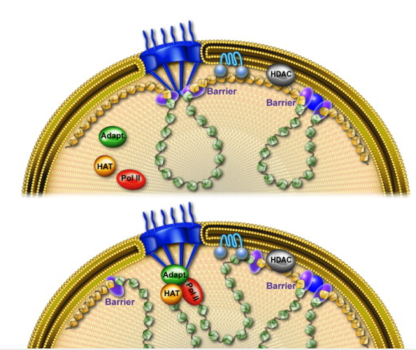
Three different organizations of this protein, some parallel some perpendicular to the plane of the nuclear envelope.

I continued to be interested in this image of a guinea pig alveolar type Ii cell nucleus, just off to the side which had a couple areas that looked for all the world as if they were part of some coiling of (coiling of the 30 nm coil) and maybe even linked together in a similar fasion as the nucleosomes themselves, but maybe packages of 3 or 4 nucleosomes with a linking region that looked to be consistent as well. Video is primitive, but it does give relevancy to the big-beads (to be distinguished from the more commonly used “beads” on a string for nucleosomally wound DNA. I really have scoured the internet and science articles to find another who has seen this kind of arrangement of chromatin at the periphery of the nucleus…. I have not found much. Nucleosomes are typically NOT as large as these one-step-further- wound DNA. Video is here:
Here is an awesome picture (diagram) which i found after making my short video…. the link to the diagram and those who made it is here. www.cell.com/molecular-cell/fulltext/S1097-2765%2810%2900172-3