Monthly Archives: February 2016

Inner nuclear membrane: do loops represent acitive DNA replication or RNA processing?

Typically the outer nuclear membrane of mammalian cells has ribosomes attached to it, the inner nuclear membrane has lamin, which appears as a band on that inside membrane. The two sides of the nuclear membrane connect at nuclear pore complexes (not seen in this tiny portion of the nuclear inner and outer nuclear membranes of a guinea pig type II cell (separated by a light band of intracisternal highly organized protein which also can fill cisternae of the rough endoplasmic reticulum in this species under some circumstances)  – see other posts in February 2016 regarding that protein.

The inner nuclear membrane, its integral membrane proteins, plus the lamins (the latter lacing the nuclear pore complexes together) participate in DNA replication and RNA processing along the heterochromain – euchromatin interface.

While working on describing the protein contents of the space between inner and outer nuclear membranes  I ran onto this really wonderful electron micrograph of a tiny portion of an of the edge of a nucleus where there were very apparent  “loops” — in the nuclear structure — maybe indicative of some sort of DNA or RNA activity (looking distinctly like beads on a string). This is a configuration which matches several diagrams found online for active (green) and inactive (orange) sites for DNA synthesis (or maybe RNA processing?) within the nucleus. I couldn’t resist trying to highlight this structure in color — at least as I saw it.

inner_nuclear_membrane_organization

Intracisternal protein: part of surfactant?

The intracisternal body shows up in the RER (the membrane of the RER is the dark boundary of the central profile and there is also some bounded by the outer nuclear membrane) but still not in sufficient detail to be absolutely sure how many bands are between the major divisions. I have highlighted these in cyan, maybe you can figure out how many are there, I think it might be 7 including both sides of the major bands (of which there are 5 in this profile of RER.

9881_17086_gp_crop_highlighted

Transmission electron micrographs of intracisternal protein in type II cells of a guinea pig – short video

I have added additional images and a pretty high magnification view of an intracisternal protein body in the type II alveolar cell of a guinea pig (Animal 301, aged, Hartley) which was used as a control in some exposure studies.  I think it seems pretty clear (that is a cya statement) that there are two ribosomes positioned just inside the dominant bands in these highly structured protein inclusions (see the last part of this video on youTube).  The dominant bands are spaced just about 0.1 microns apart.   HERE is the link to youtube.

The journal article where the cisternal body is discussed in detail is an “oldie” published in the Journal of Ultrastructure and Molecular Structure Research,  95: 131-141, 1986.

Intracisternal body in the type II cell of a control guinea pig (aged)

Working to determine whether there is an order to the ribosomes at the growing ends of the intracisternal proteins that are found in some aged animals of this and other species, I pseudocolored another transmission electron micrograph, intracisternal protein bodies are purple, and like the other images, the ribosomesa re cyan, and the lamellar bodies in the type II pneumocytes are gold-orange colored.  It seems like there might be about two ribosomes on either side of the scalloped growin end, just before the darkest of the bands running perpendicular to the growing end.  I can provide links to images that are very high resolution (26 megs) if you ask.9831_17084_gp_1_colored


Mix of science and art and emotion using polymer clay

This is an awesome testament to the power of the creative mind, mixing what one knows about biology, anatomy, physiology, physics, math, chemistry, etc etc with the need to find resolution, meaning, and beauty, release from emotional pain and also sometimes bursting with joy. We all know the lump of muscle in our chests is really a pretty ugly thing that could find its way into the meat shop and look very much like a cut of flank steak or roast. But in fact it has, from the beginning of time, been the target of our emotions and our pain and primitive man knew well that of all the organs (except what’s above the neck) this one is most vital. It quickly became the target for destruction and revenge, but also the target of love (you know… the heart ache of loss is felt right there in ones chest where the heart sits) and mourning, and excitement (increased heart rate) or peace and stillness (bradycardia). It has been symbolized and refined and iconified, immortalized, characterized, made into chocolate, paintings, lollipops, put into quilt patterns, woven rugs, appliqued, iron-ed on, embossed, tiled, stitched, politicized, and on and on, and turned into a national celebration for the entire month of February. Ha ha… we love hearts, hearts can love.
So here is a very creative “heart” which has all the earmarks of someone who loves a lot, and also knows the feeling of loss. I think it is a great statement of what the “heart” can know. I hope this woman doesn’t mind my bragging on her polymer clay vision, it is awesome. A link to her post is below.
awesome_mixhttps://www.facebook.com/photo.php?fbid=10206113713074383&set=g.318924864981444&type=1&theater

Guinea pig alveolar type II cell: protein structures within the RER

I am still trying to figure out what the ratio of ribosomes on the elongating end of these RER structures is. I think that the ribosomes appear to be staggered over the odd number of inner bands.  Here is a jpg of a type II cell from a guinea pig (aged) which actually has a huge number of separate RER profiles stuffed with this protein (which I have pseudocolored cyan) and a few mitochondrial profiles (pseudocolored purple) a couple of lamellar bodies (kind of a gold color) and again, ribosomes here and there and plentiful at that, between and at the elongating ends of the profiles of RER.

9092a_17087_gp_301_lo_res

Short animated gif: guinea pig alveolar type II cell intracisternal paracrystalline bodies.

This is just a quick fun pseudo colored electron micrograph disection of a corner of a type II alveolar cell (pneumocyte) from an aged guinea pig which was part of an inhalation study many many years ago (circa 1980s). These orderly contents of the rough endoplasmic reticulum have fascinated me for that many years and I have yet to see a good explanation for what they might be. I have seen similar inclusions in type II cells in ferret, mongrel dog and even a few in monkey.

intracisternal_body_guinea_pig_type_II_cell-1

Amazing what a little nut can do: amine sensitivity and cachews

It is just one of those “life” things, we come to the world with a set of genes and epigenitic changes, none of which is chosen and about most we are completely ignorant. Even after years of searching for what “keeps me restless at night” preventing a good night sleep I have found yet another “natural” and even healthful food which is on the list of “do not eat after 2 pm”.

It is a general tendency to think of foods that are canned or bottled or commercial to be deliberately spiked with amines (yes MSG by the boat load but other sneaky versions too) , but surprising little facts indicate that  aging and fermenting foods that are generally low in amines increases their content.

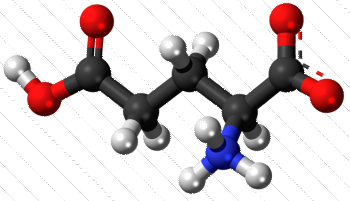
The physical and neurological impact of amines on some individuals (self included) is profound. I don’t believe as a child I understood why I wanted to jump and hop and run around, or why before trying to go to sleep at night I would feel tingly, itchy, and even get out of bed onto the floor to do sit ups to try to level out my feelings.  While I had a diligent mother, and I don’t blame her, I marvel that there was no communication about what was a bothersome physical symptom and anyone with knowledge of biology. C5H9NO4, that is five carbon atoms, nine hydrogen, one nitrogen and four oxygen atoms. The nitrogen and three oxygen atoms form the  – amine part. Wikipedia has a great post letting us know with abundant clarity that glutamine is excitatory and has other functions in memory, learning and cognitive function. So lots and lots has been written, available all over the internet, some posts are relatively scientific (I like wikipedia) and others are spooky and bloated.  glutamic_acid
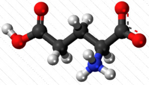
Your body typically can make all the glutamine it needs, and glutamine in most food groups abound.

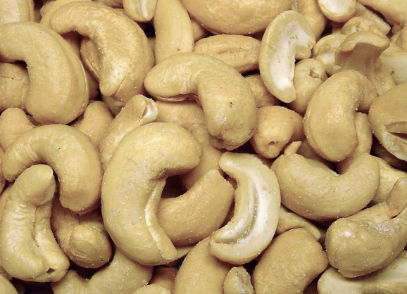
But for me, the real proof is in how I sleep at night…. which brings us to the next point — cashews. Cashews are rich in essential nutrients, vitamins and  MSGminerals, and can be part of a healthful diet. But, like peanuts, cashews are pretty high in glutamine. I am not advocating for avoiding cashews but for me, eating a handful before bedtime might be enough to trip the “no sleep” switch for you as it does for me.  It took me about 4 nights of tossing and turning to recognize that one large handful of my favorite home roasted mixture of cashews and slivered blanched almonds was sufficient to cause the symptoms.  Certainly not the most scashewsevere sleeplessness I have experienced…. ha ha… that came after eating McDonalds fries at 7 pm one evening. I will never ever do that again.  This is a matter of “dose”, but still annoying enough to remember, especially in light of the fact that our individual capacities to utililze this abundant nutrient (which over the millions of eons directed the use of same in most living sentient creatures) can also be affected by individual differences in genes and mutations therein.  This is gene environment interaction.

http://www.millhousemedical.co.nz/files/docs/factsheet_7_amines_in_foods.pdf

https://en.wikipedia.org/wiki/Glutamic_acid

Animated gif: ribosomes, grid, intracisternal protein in ferret type II alveolar pneumocytes

The paracrystal inclusion in the ferret (and guinea pig and mongrel dog) rough endoplasmic reticulum of the type II alveolar pneumocyte is really a fun structure. I have been examining old transmission electron micrographs in an effort to uncover just a little more about its structure and occurrence. I don’t think I realized back in 1986 when i published a peer review manuscript on this structure that it could be found in cross section to look like a tube, with a central point and concentric rings (see post several days ago). So looking for more confirmation on that aspect of the body I decided to make this short animated gif which shows the original electron micrograph and fades into a pseudocolored and enhanced electron micrograph where the ribosomes that feed the intracisternal protein body are highlighted in cyan and the intersection of two perpendicular alignments I have used the “burn” tool in photoshop to enhance. NO structures were deleted, this is just processing of the original micrograph.  intracisternal_body_ferret_type_II_cell

Organized protein structure in ferret type II pneumocyte (alveolar cell) rough endoplasmic reticulum

For several decades I have been wanting to study these intracisternal protein bodies, since they are spectacularly organized, seem to grow from two ends, and have large ribosome-protein complexes at the growing end and can sometimes be seen in cross section as concentric rings with a central point.  I pseudo-colored this ferret type II cell in with colors. It is from a collection of photomicrographs which depict these bodies and an especially interesting and organized sample.  High resolution images are available on two separate image libraries, but here is a thumbnail image.

ferret_type_II_cell_control