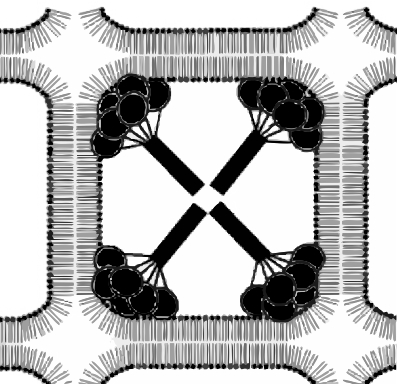
There are 12 or so images in this video which were derived from, initially, then processed with photoshop, sized with corelDRAW, animated with Swishmax 4 to reveal MORE than the 4 dense areas shown in most diagrams of surfactant protein A and its organization within tubular myelin. Hopefully these images help in detailing the areas of where the carbohydrate recognition domains of surfactant protein A might reside in a lattice of tubular myelin in the alveolar spaces of the lung – derived from diffraction images. Proposed diagrams (such as those proposed by Nades Palaniyar et al, seem to me not to fit these images very well. The final diagram published by Nades Palaniyar et al as the arrangement of four octadecamers of surfactant protein A in the corners of a more or less square lipid bilayer configuration for tubular myelin (which ALMOST matches that of countless electron microscopic studies of tubular myelin) falls just a little short of a good match. There is a space between the carbohydrate globular areas and the bounding lipid bilayer in most electron micrographs for which no account is made in thediagram found in their publication in Comparative Biochemistry and Physiology Part A, in 2001, nor is there mention of the many other dense areas in the surfactant protein A images from a previous publication (diffraction) from the same group. So i am trying to figure out what might be just a little different than displayed in their diagram. In this video composite of their diffraction images, I was able to see that not just four prominent “corner” areas of dark appear (presumably representing the globular carbohydrate recognition domains – 18 in all) of surfactant protein A, but that there are in fact 5 large areas, not 4, one of which is central, and at least 8 smaller dense areas most probably rotated 45 degrees, which should indicate some smaller structures are involved in the final organization of surfactant protein A in tubular myelin.
All this comes as a side issue of trying to figure out the periodicity of the semi-crystalline layering of an over-abundant protein in the cytoplasm of type II alveolar cells in several mammalian species, which i think is probably surfact protein A.
Their original diagram is used here (i might add – without permission) vectorized. And just an aside, not all the fatty acids in their diagram should be straight chains… there are considerably more unsaturated fatty acids in the surfactant lipid mix than shown.