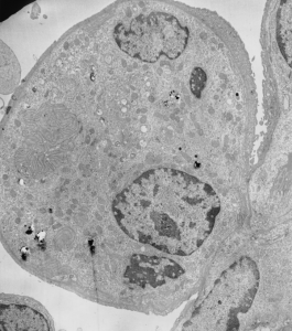
In a study by the late Dr. David Warshawsky, I took this electron micrograph of rabbit lung after exposure to Fe2O3 (you can see the iron deposits within this cell, as they ruined the sectioning knife (not so LOL). I really don’t know what cell type this is but wanted to post the micrograph in case it was of value to anyone. I think it is probably an alveolar macrophage, but found the highly convoluted plasmalemmas found as clusters in several splaces around the periphery of this cell. I figured it was significant, but didn’t what it represented. Treatment would be the first big clue. 4447_17793_rabbit_DBC and Fe2O3 .
