Category Archives: Environment and contaminants

These blogs are about our interface with our environment, mostly the food and drink that is our greatest environmental interface, in my educated, anatomist’s opinion.

Metal? cristae inclusions in mitochondria

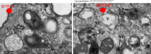
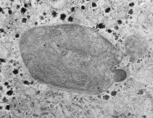
I found a paper that showed a TEM of a dense round intracrista inclusion that had some similarities to what is seen in the Gclcwc/ii hepatocyte specific KO mouse at 50 post natal days.  Paper is Characterization of Intracellular Inclusions in the Urothelium of Mice Exposed to Inorganic Arsenic Toxicological Sciences 137(1), 2013 by Puttappa Dodmane et al. I cut and pasted part of one of their electron micrographs of a mitochondrion with such an intracristae inclusion (right side of picture) next to what is seen in the KO mouse (my picture, left side of image).


YES: GRAVITY is part of the environment

How awesome is this study?  Ha ha…. having been in environmental research for 4 decades, never once did i consider looking at cells without the affect of gravity, and not to make a really bad pun, the gravity of the loss of gravity. FIND THE ARTICLE HERE. This group presents some great micrographs of the nucleolus….  I cant wait to see if the non-bilateral division of the nucleolus in one of their figures can be expanded to the very definite bilateral organization of the nucleus in general which is seen upon preparation for mitosis.

Kudos to Flying Pig Marathon recycle – repurpose – reuse efforts

THIS IS THE KIND OF INFO ThAT WE ALL NEED TO SEE DAILY
Reduce, Reuse and Running

Thanks to the incredible help of all participants, the Green Team volunteers, and staff, the 2017 Flying Pig Marathon weekend of events diverted a total of 78% of waste, exceeding our 2017 goal of 75%. We were able to successfully reduce our carbon footprint in numerous ways, including recycling heatsheets into Trex decking and railing products, recycling food wrappers through TerraCycle, recycling unused medals and participate bibs, and repurposing medal ribbons as lanyards or recycled through Goodwill’s textile recycling program. Banners, discarded clothing at the start, and leftover food were all donated to local charities. By offering carpool parking, we reduced the number of cars on the road by 1,194. We also offset part of our carbon footprint by partnering with Taking Root in planting 5 trees along the Pig marathon course.

The Flying Pig Marathon is excited to announce a partnership with Melink Corporation of Milford as the “Official Sustainability Sponsor” of the 2018 marathon weekend.

“We are thrilled to have Melink join the Flying Pig in our sustainability efforts,” said Iris Simpson Bush, Executive Director. “The Pig and its year-long events have made sustainability a prime focus for a decade now, and Melink will only help continue our efforts to preserve the environment.”

Iodoperfluorocarbons

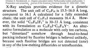
I found a very old article, portion below I did not retype but added the reference at the top, which mentioned briefly what I think is perfluorodecyl iodide (1-iodoperfluorodecane). I read this to mean that it is crystalline in nature and that the molecule has dimensions which might indicated that it has dimerized.  If I knew what a “unit cell” was I might be able to relate it to the size (which is given here in Angstrom units) of 15.5 to 16,  I might be able to figure out whether the periodicity found at the long ends of the IPFD crystals in vivo  (within lysosomal structures) and the round organization seen in some images have relevance to that dimension.

I found a more current article which seems to rehash some of what this early article by Rondesvedt mentioned in 1969. I like that he mentions that iodoperfluoroalkanes are bio-persistent, and that fits with the “mouse emulsion infusion” data that I see.  That second publication is here.  But there is no way without learning chemistry I can figure it out.

And in terms of environmental impact of PFC, in particular PFOS and PFOA and other perfluorochemicals in wide-spread usage, this is a nice article to read.

from which is a nice quote “Since the first reports revealing the widespread global occurrence of PFOS in wildlife (Giesy and Kannan 2001) and the frequent detection of PFASs in human blood (Hansen et al. 2001) were published a decade ago, the scientific literature on the environmental and toxicological aspects of PFASs has burgeoned rapidly, and the rate of publication currently exceeds 400 articles per year. In the existing body of literature, including governmental reports, authors have created terminology, names, and acronyms to describe these substances. Unfortunately, inconsistencies have inevitably arisen between various groups of authors. In the absence of any concerted effort between scientists to agree on a common terminology to designate the substances, a given compound has often been denoted by a variety of different names and acronyms, or a given acronym has been used to represent different substances. In addition, names to describe broad groups of substances have proliferated that in some instances mistakenly portray substances that are very different from one another as being the same. As a result, the scientific literature for these substances has at times become confusing. There is a need for harmonized terminology, names, and acronyms that clearly and specifically describe PFASs.” showing how difficult it is to read the literature on these chemicals.

I had to look up the words “unit cell” not being a chemist I had no clue what that meant… so here is a definition — A unit cell is the most basic and least volume consuming repeating structure of any solid. It is used to visually simplify the crystalline patterns solids arrange themselves in. When the unit cell repeats itself, the network is called a lattice.

Inert and partially inert particles in lysosomes

Here is something that might be close to looking at the endosome-to-lysosome pathway taken by perfluorochemical droplets in experiments either of liquid breathing or infusion of perfluorochemical-based blood substitutes.  I this particular article (Zhou et al, Int J. Nanomedicine, 2010 — might be a good journal to submit the PFC papers upcoming).  It is nice to see that the endosomes look a little like the dense, highly enzyme filled, MVB/LE and lysosomes seen in macrophages and other inclusions in cells exposed to PFC.  I am excerpting and editing one of their TEMs… without permission, but giving them credit, to make this parallel. Particles (Poly(d, l-lactide) (PLA) and poly-d, l-lactide-poly(ethylene glycol) (PELA) with PEG weight ratios of 10%, 20% 30%? I think this is all) don’t show a two phase morphology with their SEM images, but clearly with TEM there is a dense core and a lighter outer area. This is in total contrast to the “footprint” of PFC, in the image on the left which is basically “completely electron lucent”. But there are some similarities in the lysosome (called that just for simplicity) in each of the two images, though the PFC lysosomes (on the left) really are densely packed with enzymes, maybe even more than the nanoparticles (on the right). SEM of nanoparticles, see inset on the right.

MVB/LE/PFC bodies within alveolar macrophages of liquid-breathed mice

In this case the liquid which the mouse breathed for 3 hours and allowed to recover for 17 days was E2.  Thank goodness for good record keeping otherwise I would have forgotten what E2 stood for. Here is what the notations say (from back when Leland Clark Jr. was keeping tabs on all documents and publications.  E2 (made by Du Pont) O2 solubility ml/100ml at 25oC 55.7, vapor pressure 56mmHg at 37.5oC, viscosity 0.6 at 25oC, boiling point at 104.4oC, density gm/ml 1.656 at 25oC, surface tension 12.9 dynes/cm at 25oC.E2 liquid for breathing perfluorochemical molecular
There were a bunch of MVB/LE/PFC organelles in these macrophages that I began to look more closely at the parallel leaflet patterns that were showing up at higher magnification.

Altogether there were 29 mice that breathed E2, it much have been a satisfactory fluid or there would not be a dozen or more mice that survived from 5 days to 449 days after 3 hr of breathing E2.

The electron micrograph below was one such body, I don’t think it could clearly be labeled a multivesicular body or a late endosome, but there are E2 droplets involved in its organization.  I don’t believe there is any outer nuclear membrane association, but there are membranes with different distances apart which are clearly organized (actually a parallel disorganization – if such a thing can (and it does visually here) exist).  So I sensed that the distance between the leaflets of these membranes was different within the MVB/LE/PFC thingie and in the membranes arranged just outside the MVB/LE/PFC structure and they were different. The latter look to be about separated at 22nm n=28, x=0.053+0.0014  (mm, on the micrograph translated to close to 22nm in actuality) , and I also measured couple of dozen distances between parallel pairs within the MVB/LE/PFC structure itself and it was closer to 28nm (n=31). When the long string of measurements were subjected to a t-test (remember this is an N of ONE macrophage (there will be others) so it is not really saying much) there was an highly significant (p=0.0001) difference between the mean and SEM of the two (color coded) groups. Pictures below show how the lines and data shaped up. White dotted line outlines the two areas sampled for leaflet separation.

Figure below that shows actual sites of measurement and place of measurement for each group.

liquid breathing electron microscopy mouse lung E2

liquid breathing electron microscopy mouse lung E2In another macrophage –another micrograph — another block same E2 treatment and duration, a similar type of inclusion is seen, but this one did not display the linear qualities of the MVB/LE/PFC as above.  There are two droplets of presumptive E2 in this image, they are middle right and upper left.  Whether the lucent area in this inclusion is E2 is anybody’s guess.

Love in any language

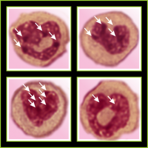
Totally unnecessary post of bone marrow nuclei: but what it says very clearly is that symmetry prevails, may not be the symmetry we expect from a set of identical chromosomes all lined up in on-off status, both sides the same (X chromosome or barr body excepted).  I see here a great symmetry, right to left mirror, top to bottom asymmetry, pointy at the bottom because this is an area where the nuclear membrane might just get segmented into a thin band between two bottom areas of chromatin…. going from what might appear to be a point, to two cheeks.

I am thinking about writing up an editorial on this symmetry…. make the cover submission first, always my motto.  Each of the hearts below is from bone marrow from an hypoplastic animal (CyP1a1null) either given benzo(a)pyrene or control diet.  These particular nuclei are not yet hypersegmented…  just fun examples of symmetry, which i have outlined in an identical micrograph below this.

four bone marrow nuclei polys chromatin condensation symmetryfour bone marrow nuclei polys chromatin condensationSo there is symmetry, it is bilateral in one dimension, but will take some thinking to see how to describe a third dimension in symmetry.

Because the chromosome territories likely (per published data) kind of interdigitate, most assuredly, at many sites along each chromosome for functional reasons) the symmetry will be highly simplified in light micrographs at best.

All this discussion could lead to a place to examine the chromatin patterns in the X (barr body) in neutrophils where they are hanging on by a nuclear membrane thread – so to speak) and other parts of the neutrophil nucleus where transcriptional activites are going on.

Electron micrograph of an hepatocyte (gd 20 fetal liver)

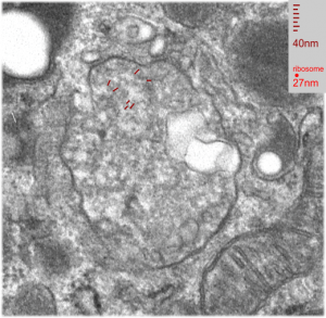
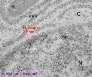
From a study in the 1980s looking at exposure of pregnant dams to CO or dichloromethane by inhalation, here is a kind of “pristine” hepatocyte from ad gd 20 fetal rat. The nuclear pores are numerous but not really that distinct, and the RER is nice and flat but also has a dense content, and the mitochondria have cristi that are not completely flattened and there is not a lot of it, and very little SER that I can find and this cell is adjacent to two hematopoietic cells, no desmosomes between. M=mitochondrion, C=cytoploasmic side, N=nucleoplasmic side arrows point to condensed (closed) and euchromatin (open). Ribosome can generate a relative size comparison for other structures, being about 27 nm in diameter (red dots), the nuclear fibrils just at the nuclear pore (almost dead center in the micrograph and within the black dotted lines) about 30 nm (black) and perichromatin granule in purple. The project, as I recall, did not uncover any differences between the exposed and non exposed animals, and in this case, the hepatocyte looks nice.

Mitochondrial cristi: what is this ordered protein

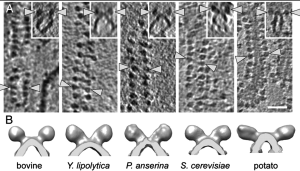
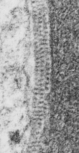
Mitochondria from vinyl chloride exposed animals have been posted on this blog before. HERE and HERE. I have made the assumption (no proof what-so-ever) that this just seems a perfect match for ATPsynthase.  Here is another example of such a mitochondrion, also showing the highly organized repeating pattern found in the intra-cristal-space of mitochondria from other liver electron micrographs from guinea pigs treated with vinyl chloride.  Amazing similarity. THis waits for someone else to confirm, but in the meantime when I see the images, I will post. Someone out there will know.  So here is a mitochondrion with such a cristi, and below that is someone elses micrograph (publication site linked) which shows the spiral repeating structure of ATPsynthase.


REF
One of their figures (not pictured here) shows about 16 dimers per 200 nm = 1 per 12.5 nm. When I measured the ribosome at 27 nm in my image (to the left) of intra-cristi structures, then there were about 19 densities per 200 nm.  So the numbers don’t add up too well.  Particularly with one of their figures copied above where their measurement (white bar in the figures and insets above) is noted in their publication to be 50 nm. This is quite far off what I see in the intra-cristi space in the figure to the left. It also seems clear that they have a propensity to be found in these groups between the outer and inner mitochondrial membranes and less frequently on the inner cristi.

Polyethylene glycol, propylene glycol, ethylene glycol, glycerol: people! wake up!

Humectants, cosmetics, antifreeze, frostings, hair products, but not good for some animals.

I don’t have to be scared to death more than about once to make a change in my behavior.  Go back about 6 years, when i had 3 dogs, all very different. One a yellow lab mix, one a husky mix, one a black brown and white generic mix.  I purchased some rawhide chews from a national chain pet store, one afternoon, and fed one to each dog.  About 11 at night, one dog was throwing up blood, the other in huge gastric distress with diarrhea and vomiting.  Hello…  this was not coincidence. The vet bill was about 300 dollars. I took the treats back to the store, the disavowed any relationship.  I never bought another chew (and assumed it was part of the melamine scandal, in retrospect, maybe it was a glycol).

January 21 2017, I visited that same pet store (under different ownership now, bought out by PetCo), and I purchased what I THOUGHT was a bleached bone. I got home and it had “peanut butter flavor” some gooey stuff in it… I knew better, but I scraped it all out, and busted the bone into pieces (sanding down the edges a tiny bit so they weren’t sharp) and fed to my two current dogs (one weimaraner and one black brown white generic mix).  One hour later, the weimaraner is kicking and jumping around then on the ground excessive salivation, in obvious gastric distress (or perhaps a petit mal seizues), i try to comfort him, we head off for the emergency room vet, where we wait for 45 minutes.  Lucky me, he recovered sufficiently to be walking around and curious and no longer in any distress (at least by outward appearance). So we left the vet, he has since been fine.  BUT I AM NOT FINE.

Next morning i called PetCo in Cincinnati and asked them to read the ingredients that match the UPC on my receipt…  fourth or fifth ingredient (AFTER WATER no less) is propylene glycol.  My weimaraner has the agouti gene, he doesn’t have the same panel of liver enzymes that other dogs do, there is much variation in this, and the human food and pet food industries are completely “clueless” about genetics.  There should be NOTHING in food that the sensitive population of individuals should not be warned of.  OF course there are food allergies, for humans and pets, but they are lacing products with non-nutritional ingredients, unnecessarily exposing humans and pets and the only reason is PROFIT.  Shameful.

The FDA claims that propylene glycol is GRAS  (generally regarded as safe)  TO ME THIS MEANS NOTHING MORE THAN — WE ARE NOT GOING TO TACKLE BIG FOOD INDUSTRIES — unless we have to.  After all, they fund us.  So sad.

Here is a newsletter so long ago about the glycols, people died. People as in human beings. Check your meds – labels.Interface_Issue_32I don’t know about you….  but it is crystal clear to me: READ THE LABEL, IF YOU CANT PRONOUNCE IT (by the way, the clerk in PetCo could not pronounce tocopherol (yes yes i know that is vitamin E… no complaints)… ha ha..let alone propylene glycol — so the people who sell this stuff are not the best sources of information. )

Here are some sites to visit.  THE BOTTOM line is NOT that all things we cant pronounce are bad, but there is “risk” in every environmental “chemical”, some naturally occurring, some artificial, some worse than others, some so toxic that nanogram quantities can end life.  The BOTTOM line is — think about WHY an additive is present, decide if it is necessary for food taste, nutrition, or if it is there for profit and shelf life.  Always recognize that we are not all created equally, except under god, but under biology all 7 billion, and counting, are unique). Find your (and your pets) uniquenesses, and be open to observation, be informed (the internet is only a good place for information if one is willing to weed out the hype), and sometimes just restrain yourself from using products full of additives.  It is dangerous enough to eat what nature has concocted by evolution in the last 14 billion years, let alone what man adds for shelf-life.

SCIENCE DIRECT

Food_safety_news_logo

BussFeed_logo


And from our own betty crocker…. a cake with propylene glycol just behind oil in the ingredient list… ha ha

betty_crocker_logo


So here is my theory:  bone is dry, peanut butter flavor gew is wet, polyethylene glycol goes to the dry bone – after all it is used to “soften and hydrate”, and is present there even though I scrape out the peanut butter flavor stuff, in just sufficient quantities to cause gastric upset.  I rule out the peanut butter allergy, because the dog has been fed natural peanut butter many times without incident.  Of course the peanut butter flavor gew is not really peanut butter.  I don’t know if the sugars, salt also seep into the bone from the filling.  Probably.