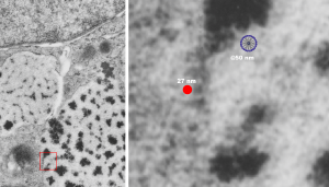
Here is a comparison, a diagram of what might be an SP-A fuzzy ball at something like 47-50 nm, and a nearby ribosome @27 or so nm. The image on left, shows inset box from the granule in this alveolar type II cell from which the inset is derived. My diagram isn’t exact, but it might be close. These images are from guinea pig (now famous) 301.
