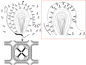
An article by Palaniyar N, Ikegami M, Korfhagen T, Whitsett J, McCormack FX. 2001 entitled Domains of surfactant protein A that affect protein oligomerization, lipid structure and surface tension, published in Comparative Biochemistry and Physiology 129:109-127, presents a diagram of how the 18-mer of SP-A might integrate itself into the outer lamellae of lamellar bodies in alveolar type II cells. I did notice an inconsistency, perhaps, in the diagrams. The bottom diagram (left bottom) shows the SP-A molecules with their N terminals all pointing inside, or towards each other in the center of these tubular myelin structures (the diagram is a 2D model of a 3D structure, hence “tubular”). In the granule found in alveolar type II cells which I have been trying to describe morphologically for several years has a layering pattern that suggests that the carbohydrate recognition domains are actually pointing “away” from each other. This is replicated in the diagram to the left. The inconsistency comes in how the SP-A molecules are represented in the top portion of the diagrams, which I duplicated in part, and re-oriented the SP-A molecules to match the tubular myelin arrangement shown in their figure (bottom left). To me this makes more sense, and it also is consistent with the layering of the alveolar type II cell granule in guinea pit, ferret, and dog, and in addition, with the outward orientation proposed for the Birbeck granule (the C-type lectin, langerin). Comments are welcome. My edits to their diagram are in the red dotted inset to right.
