So tired of looking at this structure and trying to brainstorm about how things fit together and whether SP-A is a good protein fit for this very regular and very nice molecular alignment within the RER of alveolar type II cells of several species.
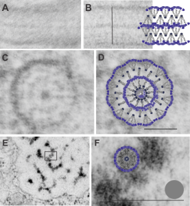
A. Single period (RER membrane on top and bottom) from ferret show outer dense layers and less dense central layer. B is also a single period of a granule from guinea pig which is not bounded by RER but by another period above and below. Less electron-density accompanies of outer dense layers occurs when adjacent periods are present. C is an end on view of a cylindrical granule from guinea pig, almost 200 nm in diameter which could be twice the single period width. The inner dense central layer would be compressed into the central density. The RER membranes are seen on the upper and left on the outer edges, and periodicities for both the less dense center layers and dense layers appear as concentric rings. D, is the same image as C, with a cylindrical model of an end-on view of the possible arrangement of SP-A molecules which could account for this structure. The number of densities in the concentric layers was used to calculate how many molecules to use for the model. Vertical lines (shown in Fig. 5 as well) became radial spokes. Bars = 100 nm. E. On rare occasions, and only in guinea pig, round and dense fuzzy-ball like structures were seen squeezed between the typical 100 nm periods. Box in E was enlarged in F to show the relative sizes of the round densities. A diagram of a potential SP-A fuzzy ball, approximately 30-40 nm in diameter approximates measurements of the spherical densities. Grey circle is ribosome size for comparison. Bars = 100 nm
