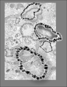
I must have really made a lot of variations for this cover submission, here is another that I modified this morning. It is a little confusing, but the cell behind is a has a large nucleus (left middle of the micrograph), which is in distress, with some indications of impending apoptosis, mainly the finely granular chromatin at the inner nuclear membrane, and the prominence of interchromatin granule clusters, and a rather large nucleolus (with prominent fibrillar centers and dense fibrillar components (in preparation for the trip down the apoptotic pathway). And on that image I superimposed a dozen or more of the images of microDNAs that Dr. J.D. Griffith gave me to organize into a cover. So the microDNAs, (of which three are enlarged bursting out in dimensional relief) that are mostly black and grey high contrast, while there are many others lesser in size that are superimposed in relief over the original transmission electron micrograph of the cell.
