Category Archives: Nuclear pores and nuclear architecture

Nuclear pores and nuclear architecture transmission electron microscopy

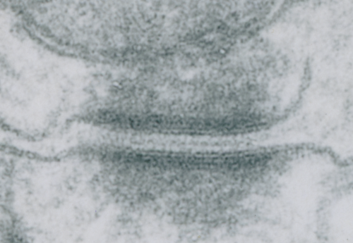
Stub tail monkey hepatocyte desomosomal-mitochondrial tether

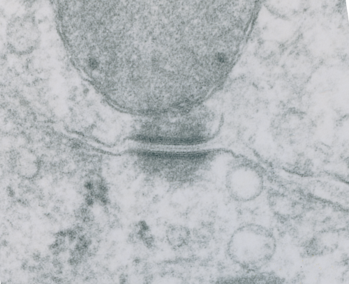
These tetherings between desmosomes and mitochondria are pretty plentiful. Here is another example of one such from a hepatocyte from a stub tail monkey. This one shows some very distinct banding…. a little more demarcated than liver, I wonder if this is just opportune sectioning or some molecular difference in the components.  Enlargement of the desmosome below top image.

I looks in this image like the plasmalemma of each cell has a very rigid separation of the two leafelets.

RNP along the inner nuclear membrane

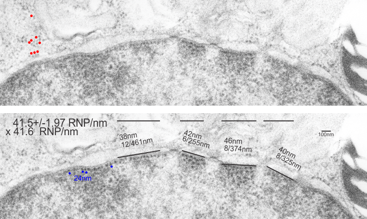
When i encounter electron micrographs where there is such obvious order i just marvel at the detail and complexity of life. Here on the inner nuclear membrane of an alveolar type II cell, in between the nuclear pores, there are little RNP particles, neatly and tidily spaced at about 41 per 100nm, and at about 24nm diameter (slightly smaller than the ribosomes from this same micrograph used at a measure of 27nm diameter. These are organizationally (that is, the RNP and inner nuclear membrane, and the ribosomes on the RER membrane) and with such similar but not identical sizes, shapes and arrangements,  that it becomes almost silly not to see an evolutionary structural relationship.

Picture on top has NOT been altered, but the identical picture on the bottom has had the distinct areas of RNP burned using photoshop just to show you what i see. Red circles are ribosome size, relative to the enlargement of the images (taken at 27nm) and blue circles are RNP granules, which are apparently closer to 24nm diameter. Point here is the rigidity of the inner nuclear membrane, and flexing of the outer nuclear membrane at sites where the ribosomes on the outer nuclear membrane are actively producing protein.

Four adjscent inter-nuclear pore distances have been measured and RNP counted. Mean+/-SEM is given at 40.5 ribosomes/100nm  and all ribosomes together with all nm is 40.6. A tangential area of the inner nuclear membrane and many RNP forming a grid like network is seen about midway-top of both micrographs, but accentuated in the lower micrograph.


@70 degree separation of homologous chromosome densities

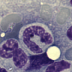
The nuclear center might not be a simple concentric center.  Why, maybe to allow for stretching and shape changing, particularly in leukocytes and migratory cells.  Seems like a pretty smart approach to something that ‘can be round’ when it is physically convenient, but is more likely to be strung out pole to pole along with the cytoplasm, or independently of the cytoplasm as well (again, leukocytes, particularly the polymorphynuclear leukocytes).  This image of a bone marrow cell from mouse has an asymmetrical shape, which is easily found, in practically any microscopic field or any bone marrow smear. Maybe like a U, but more a V really as the chromatin densities are asymmetrical in this view and in many views. Even if one takes into consideration that this is still 3D (even after spreading) and that there might be two condensed chromatin areas where there appears to be just one in this picture, the U as a whole shows a bilateral symmetry maybe in two dimensions, but not in the third dimension. This begs the question what are the symmetries in x,y and z that unfold as leukocyte nuclei become lobular.

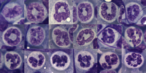
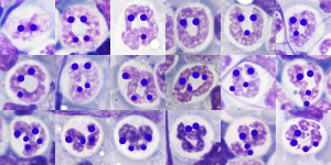
The following two images (composite markups from bone marrow smears from a single mouse. I should look up the treatment protocol, but i am pretty sure because of the number of images that it was receiving BaP.  Not all nuclei are identified as belonging to polyymorphonuclear leukocytes, but what is common is two or four sets of condensations (obviously not all can be called the Barr body chromosome drumstick) but the pattern is clear 1] bilateral left to right x axis, bilateral in the Y axis (narrow bottom two definite condensation sites at the top, and a C shape (backwards) in the z axis shown in a group of progressing nuclear lobulation, and below that, lightened images lying under dots over areas of chromatin condensation … I tried to orient the wider separation of the sister condensation points toward the top.


Arc of separation between homologous chromosomes? angular dimension?

Arc of separation (degrees of separation) , or central angle? which ever one should call it, i don’t know, between chromosome pairs in their respective chromosome territories. I see it everywhere in the FISH and other fluorescent imaging of chromosome territories. I see it in bone marrow spreads as well, clearly shown by areas of chromatin condensation of certain chromosome – or portions thereof.

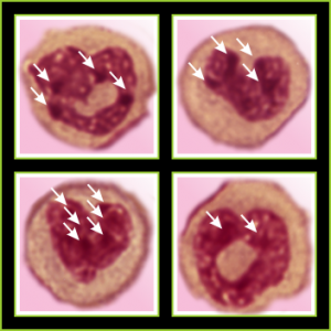
So here is a bone marrow cell which has obvious chromosomal densities, and I am presuming that these are condensed by epigenetic or other influences but there is definite symmetry (in 3 axes) and differing angular dimensions to the presumptive pairs of chromosomes. The whole nucleus is in a V shape, the point of the V being “down” here but in terms of 3D space, it may not be. It certainly represents a portion of the nucleus around which the symmetry occurs. Simetimes instead of being seen as a “v” (or heart as I have depicted it here) a U is present. and this might represent a less well condensed form of the same type of compaction, on the way to becoming a multilobular white cell.

The arc drawn between what is felt to be homologous areas of condensed chromatin, billateral symmetrical dark spots (highlighted in black on images to the right of the unretouched (but color adjusted to red) giemsa stained white cell).  The arc is influenced of course by random position of the cell as it is smooshed onto the slide during preparation as well as the spot identified as the center of the nucleus… whatever that might turn out to be, but i bet it is somewhere near the apex (bottom of the heart).  The actual physical center of the nucleus is not well defined in the literature.  So i think the concept of radial chromosome territory positions might need to be altered to account for the z axis of symmetry and the position which ultimately will be described as the nucleolar “center”… which cant be the nucleolus only, since often there are multiple nucleoli…  maybe the term center of the nucleus should be called “centers of the nucleus” and the arc of separation determined from various sites.

Love in any language

Totally unnecessary post of bone marrow nuclei: but what it says very clearly is that symmetry prevails, may not be the symmetry we expect from a set of identical chromosomes all lined up in on-off status, both sides the same (X chromosome or barr body excepted).  I see here a great symmetry, right to left mirror, top to bottom asymmetry, pointy at the bottom because this is an area where the nuclear membrane might just get segmented into a thin band between two bottom areas of chromatin…. going from what might appear to be a point, to two cheeks.

I am thinking about writing up an editorial on this symmetry…. make the cover submission first, always my motto.  Each of the hearts below is from bone marrow from an hypoplastic animal (CyP1a1null) either given benzo(a)pyrene or control diet.  These particular nuclei are not yet hypersegmented…  just fun examples of symmetry, which i have outlined in an identical micrograph below this.

four bone marrow nuclei polys chromatin condensation symmetryfour bone marrow nuclei polys chromatin condensationSo there is symmetry, it is bilateral in one dimension, but will take some thinking to see how to describe a third dimension in symmetry.

Because the chromosome territories likely (per published data) kind of interdigitate, most assuredly, at many sites along each chromosome for functional reasons) the symmetry will be highly simplified in light micrographs at best.

All this discussion could lead to a place to examine the chromatin patterns in the X (barr body) in neutrophils where they are hanging on by a nuclear membrane thread – so to speak) and other parts of the neutrophil nucleus where transcriptional activites are going on.
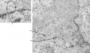
What are these round structures? too big for coated vesicles?

Here are some nice round structures and were they always near the nuclear membrane i would count them as tangentially sectioned nuclear pores since they are sized so similarly (though i have not seen any central densities like i see in nuclear pores where actual proteins are being imported or exported. There are also some profiles of membrane (with contents) that are pretty linear for golgi seen as well, and don’t quite fit that golgi iconic morphology. This is a fetal hepatocyte. Arrows point to pore and to similarly sized rounded structures that are not likely to be tangentially section pores from the nucleus shown.
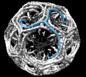
Might be a section of a clathrin coated vesicle…. when i google it, this wonderful model came up.  Sure looks (at 100nm) like it could work.  What a marvelous amount of research has provided the molecular structure (triskelion) shown by them (on wikipedia) in blue.

Nuclear pores from Gclc ko and wt mice

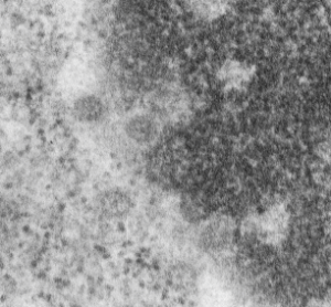
Apoptosis seems to cause a real reduction in the chromatin exclusion zone of hepatocytes.  The area is very small on either side of the nuclear pores. This would have to be functional but whether it relates to something changing about the nuclear basket portion of the nuclear pore complex, or about the chromatin, or about some other proteins that are part of the exclusion zone is up for debate. These pores are from nuclei which appear to be undergoing apoptosis, showing lots of peripheral chromatin, a little different in texture than regular condensed chromatin and an euchromatin space which is a little denser and more populated with interchromatin granule clusters than a non-apoptotic cell would be. These cells would be from livers of animals either never rescued or removed from NAC. Pore complexes are about 120nm in their widest dimension and there is another (taken from a n of 5 random pore and their exclusion zone images) another 55nm of exclusion on either side. This is in agreement with a larger selection of chromatin exclusion zones measured in both Gclc ko and 14CoS ko mice… previous posts on this site, which was 48nm and 50.9nm.

Similar small chromatin exclusion zones are found in cells in liver in eminent apoptosis in the 14CoS null mice when they are NOT rescued with NTBC.

electron microscopy nuclear pores chromatin exclusion zones in apoptosis

More nuclear pores, just randomly collected various cell types

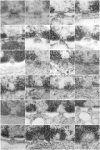
More nuclear pores, just randomly collected various cell types. The criterion that I used was simply the presence of vertical filaments (either part of the nuclear pore basket on the nuclear side or the filaments in the cytoplasm on that side. These come from lung alveolar type II cells, from hepatocytes, and from CoS14 ko mice, rescued and non-rescued, and probably a couple from Gclc conditional KO mice. These are just to give the limit of what effects random sectioning through a block of tissue can do to a round or discoid object. These pores are cut perpendicular (side views), and chromatin is always on top, cytoplasm is always below.

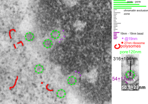
Nuclear pore measurements: pore to pore, chromatin exclusion zone, granule to granule

Three measurements taken in a series. Pore to pore, pore to to edge of chromatin (which i found is called the chromatin exclusion zone, so am sticking with that since it clearly is an active event, and may vary with cell type and cell activity) and inter-@19 nm “beads” that are seen as the organized chromatin just adjacent to the nuclear pore (the first thing seen after the chromatin exclusion zone).  This micrograph is from the same images as a previous post, which is 16027_65718_14Cos_ko 24hr no NTBC measurements.-2a. n=5,  pore to pore=316nm+104nm; n=15, chromatin exclusion zone, 50.9+23nm; n=5, 19nm to 10nm chromatin granule, 54nm+17.7nm

66 names for the devil

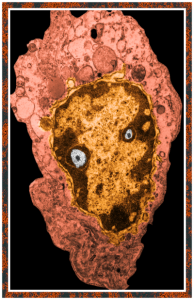
This post is just for fun, as when i was looking at some electron micrographs of the Gclc mouse liver study I found this critter, just with big eyes (one of which i added the pupil to using photoshop — my apologies) but otherwise not retouched.  The exact data are neg17894_block74138_animal706_wcii_NAC. Initially I was interested in the nucleoli, but naggingly amazed about the “devil” appearance of the nucleus as a whole.
Color was of course added using photoshop, all else remains the same. When considering what to call this character i googled “names for satan” and behold, the list of about 66 names from various cultures and groups showed up (cut and pasted from Wikipedia (thank you wikipedia) (below).
Abaddon—(Hebrew) the destroyer
Adramalech—Samarian devil
Ahpuch—Mayan devil
Ahriman—Mazdean devil
Amon—Egyptian ram-headed god of life and reproduction
Apollyon—Greek synonym for Satan, the arch fiend
Asmodeus—Hebrew devil of sensuality and luxury, originally “creature of judgment”
Astaroth—Phoenician goddess of lasciviousness, equivalent of Babylonian Ishtar
Azazel—(Hebrew) taught man to make weapons of war, introduced cosmetics
Baalberith—Canaanite Lord of the covenant who was later made a devil
Balaam—Hebrew devil of avarice and greed
Baphomet—worshipped by the Templars as symbolic of Satan
Bast—Egyptian goddess of pleasure represented by the cat
Beelzebub—(Hebrew) Lord of the Flies, taken from symbolism of the scarab
Behemoth—Hebrew personification of Satan in the form of an elephant
Beherit—Syriac name for Satan
Bilé—Celtic god of Hell
Chemosh—National god of Moabites, later a devil
Cimeries—Rides a black horse and rules Africa
Coyote—American Indian devil
Dagon—Philistine avenging devil of the sea
Damballa—Haitian Vodou serpent god
Demogorgon—Greek name of the devil, it is said should not be known to mortals
Diabolus—(Greek) “flowing downwards”
Dracula—Romanian name for devil
Emma-O—Japanese ruler of Hell
Euronymous—Greek Prince of Death
Fenriz—son of Loki, depicted as a wolf
Gorgo—dim. of Demogorgon, Greek name of the devil
Haborym—Hebrew synonym for Satan
Hecate—Greek goddess of the underworld and witchcraft
Ishtar—Babylonian goddess of fertility
Kali—(Hindu) daughter of Shiva, high priestess of the Thuggees
Lilith—Hebrew female devil, Adam’s first wife who taught him the ropes
Loki—Teutonic devil
Mammon—Aramaic god of wealth and profit
Mania—Etruscan goddess of Hell
Mantus—Etruscan god of Hell
Marduk—god of the city of Babylon
Mastema—Hebrew synonym for Satan
Melek Taus—Yezidi devil
Mephistopheles—(Greek) he who shuns the light, q. v. Faust (Greek)
Metztli—Aztec goddess of the night
Mictian—Aztec god of death
Midgard—son of Loki, depicted as a serpent
Milcom—Ammonite devil
Moloch—Phoenician and Canaanite devil
Mormo—(Greek) King of the Ghouls, consort of Hecate
Naamah—Hebrew female devil of seduction
Nergal—Babylonian god of Hades
Nihasa—American Indian devil
Nija—Polish god of the underworld
O-Yama—Japanese name for Satan
Pan—Greek god of lust, later relegated to devildom
Pluto—Greek god of the underworld
Proserpine—Greek queen of the underworld
Pwcca—Welsh name for Satan
Rimmon—Syrian devil worshipped at Damascus
Sabazios—Phrygian origin, identified with Dionysos, snake worship
Sammael—(Hebrew) “venom of God”
Samnu—Central Asian devil
Sedit—American Indian devil
Sekhmet—Egyptian goddess of vengeance
Set—Egyptian devil
Shaitan—Arabic name for Satan
Shiva—Hindu the destroyer
Supay—Inca god of the underworld
T’an-mo—Chinese counterpart to the devil, covetousness, desire
Tchort—Russian name for Satan, “black god”
Tezcatlipoca—Aztec god of Hell
Thamuz—Sumerian god who later was relegated to devildom
Thoth—Egyptian god of magic
Tunrida—Scandinavian female devil
Typhon—Greek personification of Satan
Yaotzin—Aztec god of Hell
Yen-lo-Wang—Chinese ruler of Hell