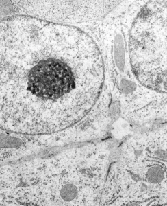
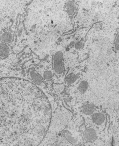
Fetal liver: electron micrograph which is pretty unremarkable. This particular image is take from a fetal rat at 20 days, but the mother had been exposed to dichloromethane during days 7- 20 of a timed pregnancy. It has a great nucleolus for sure and the mitochondria are quite nicely7 preserved, the hepatocytes themselves don’t have a lot of stacked RER, and the bile canaliculus is really well organized and the little microvilli and junctional complexes among the four hepatlcytes pi9ctures are unremarkable. There is a cell at the top of this micrograph which looks to be an hematopoietic cell (probably part of an erythroblast). (57
While I did some electron microscopy for this study I don’t believe it was ever published. The PIs were J Manson and B Hardin, and some literature does exist on their experiments.

