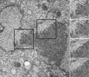
Here is kind of an interesting set of features in this clump of granular component of the nucleolus (or may just be condensed chromatin) that has a checkered appearance in some places (see checker boxes in inset micrographs to the right of the original image one showing the unretouched sections and an identical one with a checker pattern highlighted). There is a definite order to the spacing… or banding or filament pattern, something around 125 nm for the top two inset images, and for the bottom inset images, around 50 nm (which goes along with most measurements — being too big for interchromatin clusters granules (20-25nm), splicesosomes, around 30 nm, possible speckles? which are more like 50-100 nm).
