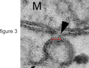
I just love it when I can see patterns in electron micrographs. I am not saying that there are not patterns in globs, and bumps and lines etc that appear as artifacts of fixation, of course they do, that is part of the nature of this particular process of inquiry. All methodologies have them, this one, that is the denaturing and folding and dis-alignment of molecules seen in aldehyde (or any) fixation, belongs to microscopy. BUT WHEN i can see repetition in an orderly pattern as in these red dots, then i think, OK there is an underlying organization of the proteins (whether by fixation made lumpy or round or whatever), there is organization. The authors that created this image have looked at mitochondrial-RER and SER interfaces and given reason to believe that there is a transport of Ca+ back and forth using SERCA 1 (sarco/endoplasmic reticulum Ca2+-ATPase 1s). They don’t mention the regular areas of protein denaturation here, but I think it would be highly possible that it relate to some aspect of the SER membrane proteins coming in close contact with outer mitochondrial membrane. I added the red dots, just below actual dots in the micrograph, highlighting the symmetry of spacing and size. Whether this relates to their fixation, or stain, their negative resolution or a structural organization on the SER i don’t know (the overall grain density of this micrograph is similar to the spot size), but it caught my attention. Their figure below is from — Leopoldo de Meis , Luisa A. Ketzer, Rodrigo Madeiro da Costa, Ivone Rosa de Andrade, Marlene Benchimol . Fusion of the Endoplasmic Reticulum and Mitochondrial Outer Membrane in Rats Brown Adipose Tissue: Activation of Thermogenesis by Ca2+. PLOS One Published: March 2, 2010