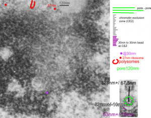
The nuclear pores are really abundant around the bounding membrane of the nucleus, especially in fetal liver cells, and other metabolically active cells. This begs the question as to whether nuclear pores can be measured in order to predict their frequency, and the width of the chromatin exclusion zone around them. The diameter of nearby ribosomes, taken at 27nm, is used as a standard, as is the diameter of the chromatin beads just inside the inner nuclear membrane, just slightly larger than the ribosome. The inter-bead distance (aka the distance between-chromatin-beads at the margin of the chromatin exclusion zone) and distance from nuclear pore to this string of chromatin “beads” of the condensed chromatin beside it has been made, along between pore distances This being an alveolar type II cell instead of an hepatocyte, didn’t really define any great differences in these three measure compared with hepatocytes. 10291_18339_ferret_1_alveolar type II cell. Mean distance between nuclear pores in 6 samples so far is approximately 332nm+/-30nm (SEM)
