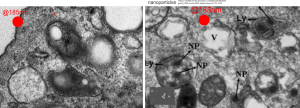
Here is something that might be close to looking at the endosome-to-lysosome pathway taken by perfluorochemical droplets in experiments either of liquid breathing or infusion of perfluorochemical-based blood substitutes. I this particular article (Zhou et al, Int J. Nanomedicine, 2010 — might be a good journal to submit the PFC papers upcoming). It is nice to see that the endosomes look a little like the dense, highly enzyme filled, MVB/LE and lysosomes seen in macrophages and other inclusions in cells exposed to PFC. I am excerpting and editing one of their TEMs… without permission, but giving them credit, to make this parallel. Particles (Poly(d, l-lactide) (PLA) and poly-d, l-lactide-poly(ethylene glycol) (PELA) with PEG weight ratios of 10%, 20% 30%? I think this is all) don’t show a two phase morphology with their SEM images, but clearly with TEM there is a dense core and a lighter outer area. This is in total contrast to the “footprint” of PFC, in the image on the left which is basically “completely electron lucent”. But there are some similarities in the lysosome (called that just for simplicity) in each of the two images, though the PFC lysosomes (on the left) really are densely packed with enzymes, maybe even more than the nanoparticles (on the right). SEM of nanoparticles, see inset on the right.
