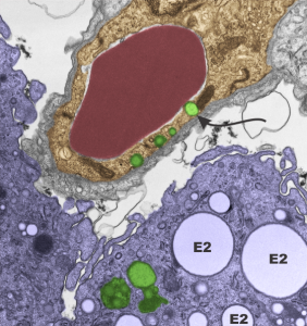
Loooooking over an old negative of lung from a mouse that breathed oxygen bubbled liquid perfluorochemical (in this case E2, which is a freon) I spotted what could be, maybe, seems almost possible, droplets of E2 in the endothelium (alveolar sac endothelium). I selected portions of the image out and pseudocolored them for easy identification: Red cell within the capillary lumen, red, endothelium orange, lysosome/or vacuoles that might be E2 and comparison with those which are in a nearby alveolar macrophage, green, and the macrophage cytoplasm is purple. There are some reasonably large E2 droplets which are probably unequivocal (is that not an odd combination of words?) with more distinct membrane boundaries, and absolutely NO fuzziness within the inclusions but also having the distinctive “black cap” of lysosomal enzymes off to a little blip on some side or other of the droplet. This is NOT seen in the endothelial inclusions…. I will look for less equivocal samples but the similarities between those and additional ones found in the macrophage is clear. 1351_4840_mouse_E2 breathing for 3 hours, 48hr recovery, capillary_endothelium and two macrophages.
