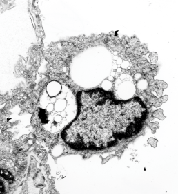
Here is a not-so-hot electron micrograph of an alveolar macrophage, but the inclusions of silicone oil are kind of interesting. By 72 hours after just one hour of breathing silicone oil, show droplet inclusions that look a lot like they are doing the Oswald ripening thing, but also havn’t stimulated quite the lysosomal enzyme thing that macrophages that have phagocytoses perfluorochemicals show. There are also bizarre areas of the lysosomes which are not typical of the PFC inclusions. So differences – noted!
One of the more interesting differences (well two) is that there is almost no discernible trilaminar membrane around the inclusions (LE/SiO droplets) and there is certainly not a double outline (one is the trilaminar membrane of the lysosome, the other the protein coat as seen on PFC droplets (see previous posts)), so those two differences in ultrastructure plus the lack of a definite enzyme overload in those particles in alveolar macrophages that contain silicone oil droplets, and the sloppy separation (or ripening) of the individual droplets of silicone oil vs PFC droplets, makes for nice contrast between the two.

Here on the left is an enlargement of silicone oil inclusions in an alveolar (1 hr breathing 72 hr recovery) macrophage and on the right, E2 inclusions 3 hr breathing 48 hour recovery (biasing the E2 in the direction of less organization, but still showing much more organization and lysosomal enzyme content than the silicone oil).