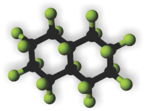
PP5 or perfluorodecalin (image to right) was used as a possible component of artificial blood, or called otherwise, blood substitute(s), but never really attained full research or understanding necessary to determine whether it was a safe substitute for blood or not. Some of the early electron microscopy, which would have benefited from a huge influx of money, early on, for microscopists to examine the effects on all cell types, in vivo and in vi tro, but too little was forthcoming to give it a thorough exam. It was clear however, that macrophages and the immune system were going to be big players, both because of the volume of the chemical that was infused or “breathed” and because of its alleged inert nature but odd surface tension properties.
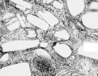
In retrospect, looking at tissues which were taken from Leland Clark Jr’s lab in the 1970s, these particles (droplets) behaved strangely, seemingly dependent upon chemical properties, and of all examined ONLY one perfluorochemical had a shape in the cell other than very very round (this was perfluorodecyliodide – looking like a crystal instead of a droplet, as seen in this image from the liver after infusion of an emulsion).
Liquid in the cell in almost always is “slightly out of round” the best examples being fat droplets. They can be oval or indented or squeezed, as well as round.. but perfluorochemical droplets are ROUND. I would think this could be attributed to their heavier than water density.
Another property (mentioned in previous posts) is that they have various capacities to be re-emulsified, at least that is what i call it, within the cell. Example from the liquid breathing mouse posted yesterday (breathing silicone oil vs breathing E2) shows that the respective macrophages respond by producing different quantities, and also likely types of lysosomal enzymes, in addition to the recycling they do of the alveolar contents (that is, in recycling surfactant and other junk in the alveolar space.
So comparing silicone oil, E2 and PP5… seen here, once again, the lysosomal responses are quite different. Silicone oil didn’t encourage lysosomal production (and surfactant recycling) as much as E2, and E2 in turn caused a lesser (though still pretty dramatic) response than PP5. Actually, from notes taken from this series of experiments (Clark never did anything twice which made it really tough to keep notes) those mice breathing PP5 did not fare well, most did not survive as long as those breathing some other of the perfluorochemicals. E2 was probably the best in this regard, just due to the sheer number of samples and micrographs, this is what I remember.
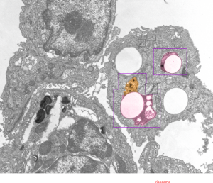
So here is an electron micrograph of an alveolar space from a mouse which breathed PPF for 1 hour and was allowed to recover for 1.5 hours. (1499_5512_PP5_1hr_1.5hr recovery).
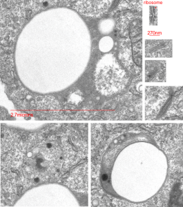
 Highly rounded droplets, with very well defined lysosomal enzymes. The two non-highlighted droplets are likely to have (as the upper left already indicates) masses of lysosomal enzymes in a different plane of section. There are some smaller, and also very tiny droplets of PP5 visible in the two highlighted LE/PFC bodies. I did find some dense areas of the ER (not likely RER, but something different) that showed an unique substructure (see the inset with three small sample images (enlarged from boxes in the top figure) showing what I noticed.

Inset is enlarged and micron marker is determined from the size of an average (27nm) ribosome, and segments of the inset are all at the same enlargement from the original.