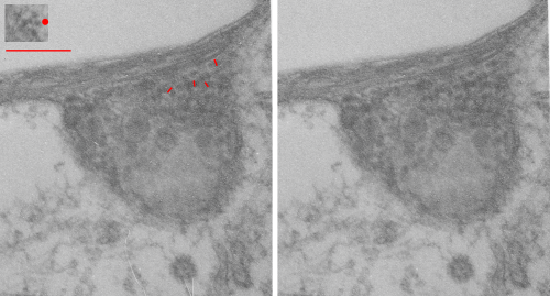
Phagolysosomes which include IPFD – 1 iodoperfluorodecane aka perfluorodecyl iodide have some interesting lysosomal enzymes.
Of these many enzymes I am thinking that there are many which are “folded” or oligomerized, that is those phagolysosomes which have IPFD show a heterogeneous substructure, zones of tube like enzymes, and areas of mostly untextured protein and then areas of unique coiled ultrastructure. Below are two electron micrographs, the one on the left unretouched scan of a print, and the one on the right retouched (though I can hardly tell the difference – the one on the right had the coiled nature of the protein burned just a tiny bit with photoshop and several pieces of lint minimized with the bandaid tool in photoshop but no data were changed). Red dot is ribosome (taken at about 27nm) and the thickness of one of the fibrils is something under 25nm… which is a little larger than i measured before at 20nm but i am probably responsible for the variation), red bar is 270nm, short red lines are measurements of the diameter of the coiled (polymer or oligomer protein), pale untextured area at the top of the micrograph is the footprint of the IPFD crystal, the whole area is mostly phagolysosome. Inset is the cluster of ribosomes used for size.
