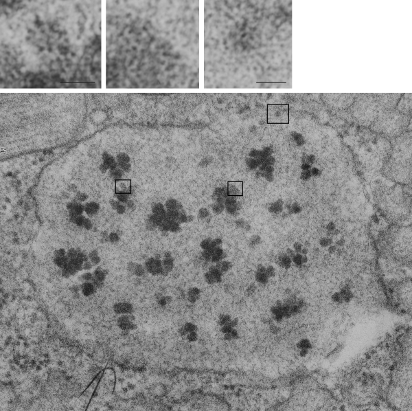
This particular profile of RER in a type II alveolar cell of a guinea pig has bothered me for at least 3.5 decades. The guinea pig is aged but otherwise untreated, i.e. wasn’t part of a experimental treatment protocol but was part of a study on toluene diisocyanate exposure. This RER profile has very large cluster inclusions within the periodicity, which is very difficult to see in this particular profile because of the tangential sectioning. I have no clue what this is, but it shows considerable substructure on enlargement of the micrograph (taken at 23,500x, with the V pole piece in an old Siemens 1A electron microscope, and the enlargement in the darkroom was 4x. Scan was 3200 ppi.
Several areas which showed a terrific hexagonal substructure were cut and pasted, a marker for their relative nm bar scales given in each, based upon a repeat measure of local (the same micrograph) ribosomes at a pretty much standard 20 nm size. Please help identify these (ha ha) (I do have comments turned off for obvious spam reasons) you will be able to contact me in other ways.