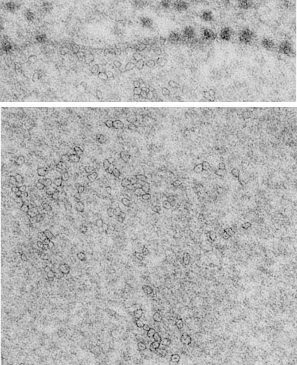
Plugging away at whether these hexagonal structures found within the RER protein accumulations in some alveolar type II cells which are found on tangential sectioning of what I have called “intracisternal bodies” are really SP-A bouquets which are splayed out with a center dot being the neck and collagen portions of that molecule. I have taken a portion of one such intracisternal body which has a small portion which is ribosome studded highlighted (using the burn tool in photoshop) a few of the hexagonal arrangements which seem to have six or so densities on the perimeter, possibly the carbohydrate recognition domain trimers of the SP-A 18-mer.
Previously posted images of those found in ferret type II alveolar cells really looked to be larger, and that is problematic. These hexagonal structures (pictured below) are from an aged, untreated, control, guinea pig and the bouquets seem more in line with the size of a 20 nm ribosome, a string of which I have included for assisting with perception of relative sizes. There is a large tangentially sectioned dark band (actually two dark bands) in the image below unaltered, enlarged with the ribosomes in which I have highlighted the hexagonal molecules (using ONLY THE BURN TOOL in photoshop) to accentuate what I see as patterning. The ultimate test of this would be to take an unrelated micrograph of an unrelated RER inclusion, at the same magnification and see if i could pick out as many hexagons per nm in it as I find in a tangential section of an intracisternal body ( ha ha–sort of pedestrian but it would be a good test).
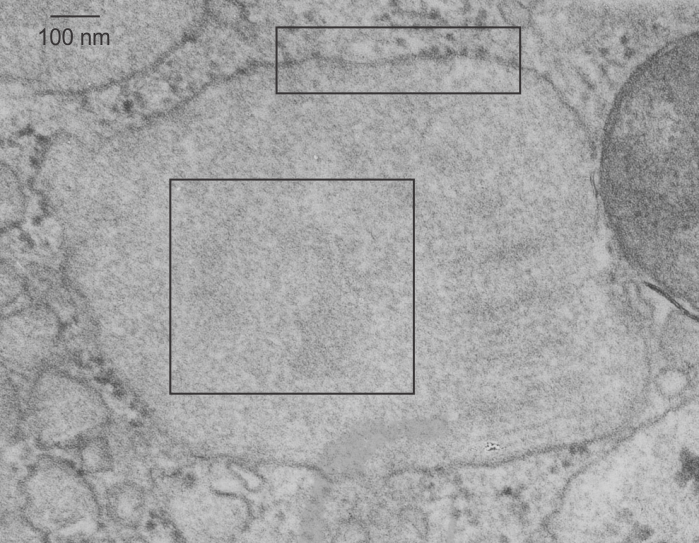
So the upper micrograph gives you a distance marker (bar=100 nm) and the upper box gives you the area of ribosomes and a portion of the intracisternal protein structure which I presume to be SP-A. The lower box gives you the identical area of as is found in the bottom panel, which has two areas of slightly darker electron density marking the tangential portions which are the dark lines of the intracisternal body periodicity. In both enlargements (boxes) I have highlighted just a few of the very many hexagons, which are hopefully the top down view of SP-A molecules.
For those of you who are curious, a mitochondrion is off to the right, and there are also portions of two large intracisternal bodies, and half a dozen or more single – periodicity intracisternal bodies as well. In the central intracisternal body on the right hand side shows the 100 nm periodicity (which on a completely perpendicular section contains anywhere from 3 – 9 bands).