I found this paper by Doreen Ashhurst on insect desmosomes which had some transmission electron microscopy of moth desmosomes which are clearly very different than those found in mammals. Brief notes. JCB 46: 421-425, 1970. It is a scanned pdf so the images are not that well preserved but from the text and images here is a set of general similarities and dissimilarities between moth and mammalian desmosomes (typical epithelial cell connections). The list begins at the most intracellular zone to intercellular space, beginning with the cytoplasmic structural proteins, the plaque itself (the outer dense plaque and inner dense plaque in mammals), the plasmalemma, and the central dense line where the cadherins hook up (at least there is a central dense line in mammalian desmosomes, seemingly not in moth desmosomes) .
| WAX MOTH | MAMMALIAN |
| 600nm | 250-300nm diam |
| oval shape | pretty much round |
| cytoskeletal protein=microtubules | cytoskeletal protein=intermediate filaments |
| MT parallel to desmosome | IF parallel to desmosome |
| lucent area by microtubules | desmoplakin molecules by IF |
| fuzzy outer plaque w periodicity | neat tidy outer plaque w periodicity |
| periodicity about 20nm | periodicity about 4nm apart |
| intercellular space about 20nm | intercellular space at desmosome @10nm |
| desmosome has an annulus | desmosome has an annulus |
| intercellular densities periodic | intercellular central dense line |
One thing seems true, the periodicities are less marked in insect (thought the X and Y configurations of what would be equivalent to the desmogleins and desmocollins are just barely seen in this photo by Ashhurst I bet they do have some relationships to mammalian desmosomes. The differning dimensions of the desmosomes between moth and mammal are really quite interesting.
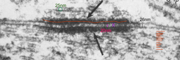
Her micrograph did not have a scale bar but I used a microtubule (with a diameter of 25nm) as the standard. Red line through desmosomal outer plaque used to measure the diameter in the long dimension (presumably) of the insect desmosome. Microtubule in the upper left used to get an estimate of overall magnification of the image (microtubule at a nominal 25nm diameter). Similarly, moth to mammal, there was a slightly reduced width (height) of the intercellular space at the central part of the desmosome, than there was at the extracellular space between the adjacent cells at the points away from the desmosome. The distance within the desmosome being smaller than the distance (height or width of the intercellular space) adjacent but which is NOT part of the desmosome. There is an annulus for the moth desmosome, just like for the mammalian desmosome.
I love that this author says…. “intermediate filaments may run parallel to the plasmalemma in mammalian desmosomes…. ” i know she saw that problem with the early interpretation…. thats fun, and she saw it way back in 1970, and the same is misjudged today. Old habits die hard, and hardly die at all.