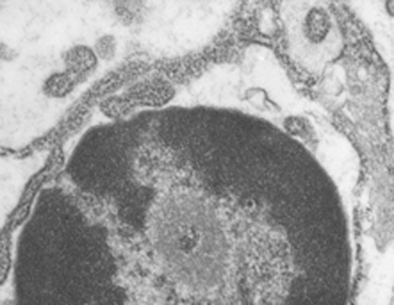
I wish i had taken a higher mag picture of this apoptotic nucleus as well as identified the cell type. I do know it is from animal #500 in a study of aged ghkaα-/- mice, this particular KO was 12 months old. It could be a parietal cell since in other image parietal cells are shown to have a “glassy protein” (stains without much texture and eosinophilic with H&E) (just recollection here). The RER is greatly, ribosomes are very closely packed (indicating the production of large amounts of possibly large protein products. Checking the fairly electron lucent contents of the RER there doesn’t seem to be any obvious oligomerization, polymerization or layering of the contents. The light areas in the perinuclear space and around is dilated RER. It could also be a zymogen secreting cell.
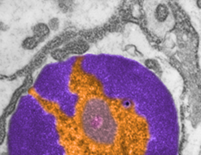
 One nuclear pore on the middle left nuclear membrane, and a part of another to the right and above that one. Packed ribosomes are in array on the RER membranes (the black and white cytoplasm pictured around the nucleus), lots of condensed chromatin (purple) which may be exaggerated by the section being somewhat tangential to the widest diameter of the nucleus, and adjacent euchromatin (gold) which in this case looks pretty dark (not just because i pseudocolored it). There is a prominent perichromatin granule just to the lower right of center (black) and interchromatin group (sometimes called nuclear speckles which to me makes no sense) is light violet. Pinkish spot in the interchromatin granule is probably just euchromatin?
