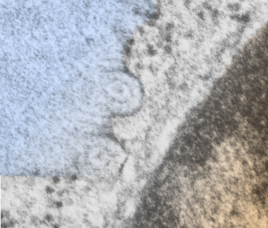
More images which pique the imagination about what is responsible for the highly organized protein in the RER of the type II alveolar cell in some mammals (in this case an aged guinea pig, control, from a previous study). I have mentioned before that there are examples of wildly converging and blending and mixing of these proteins going from a perpendicular arrangement to parallel to the long axis of the RER profile and splitting off and curving and also concentric or semilunar. This is a really nice resolution and relatively clear image of a tubular arrangement, cut in cross section, comprising a single “period” that is here seen as center dot, medium density ring and outer ring. This short video is constructed from two images, the initial image is untouched, the fade in image i have used photoshop “burn” tool to highlight the area that looks to me like it is concentric.
I added a blue color to an overlay of the actual cisternal body on the left, and a brownish overlay to the nucleus at the bottom right corner. Ribosomes are clearly found, and are especially prominent where the banding runs parallel to the RER, just above the tubular section.
Perinuclear space might have a trace of this protein in it, perpendicular to the direction of the nuclear membrane but it is quite indistinct, and there is a perichromatin granule very near the nuclear pore seen on the left hand side of this profile of the nucleus.
video is available on YouTube:
Composite image of intracisternal body (blue, nucleus, brown)
