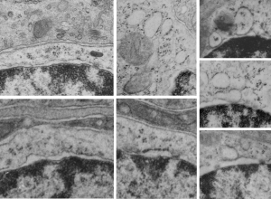
I am continuing to search for electron micrographs which have images in high resolution and magnification of the c-type lectins to compare with the intracisternal protein accumulation in type Ii cells of the lung of some species of mammal. Upon thinking about the, comparing with the fine structure of langrin in the Langerhans cell, it occurred to me that I might have a similar structure in the microvillar cell of the olfactory epithelium.
The microvillar cell has not been a topic of much research, and it has no well established function. Because of the occurrence of about 10% of most epithelia in the body to be burried in the epithelial cells proper (10% in olfactory epithelium, back skin, colon epithelium, etc) that I have examined personally it made sense to go over that set of about 250 micrographs looking specifically for RER profiles that specifically had a periodicity or layering to the protein content. Unfortunately, in all those microvillar cells examined, I did NOT find any organization to the protein contents of the RER profiles, but I did find the sparse ribosome studding on the profiles that I have seen in the type II cell of the lung.
Nothing conclusive, but just a little bit peculiar. I do wish I had seen some indication that the RER of the microvillar cell had a “granule” like the birbeck granule… ha ha…. that would have given an instant functionality to the microvillar cell. It might still be there… as not all RER protein contents show periodicity like the one in type II cells and Langerhans cells. I have not found images actually for any other collectin that speak to layering, just the one I have examined and the birbeck granule in Langerhans cells.
Here is a quick composite of some profiles of RER in the microvillar cell of the olfactory epithelium (recall, that upper layers contain the olfactory bipolar cells, the sustentacular cells (supporting) and microvillar cells (at about 10% occurrence. Profile of nucleus in the bottom sector of each image, perinuclear space and RER profiles are just above that. RER profiles are quite electron lucent, and only on e of the pictures shows any contents (and to me those are too close in shape to ribosomes to be excited about).
I would love to see any electron micrographs of MBL or other c-type lectin protein overexpressed in any type of cell.
