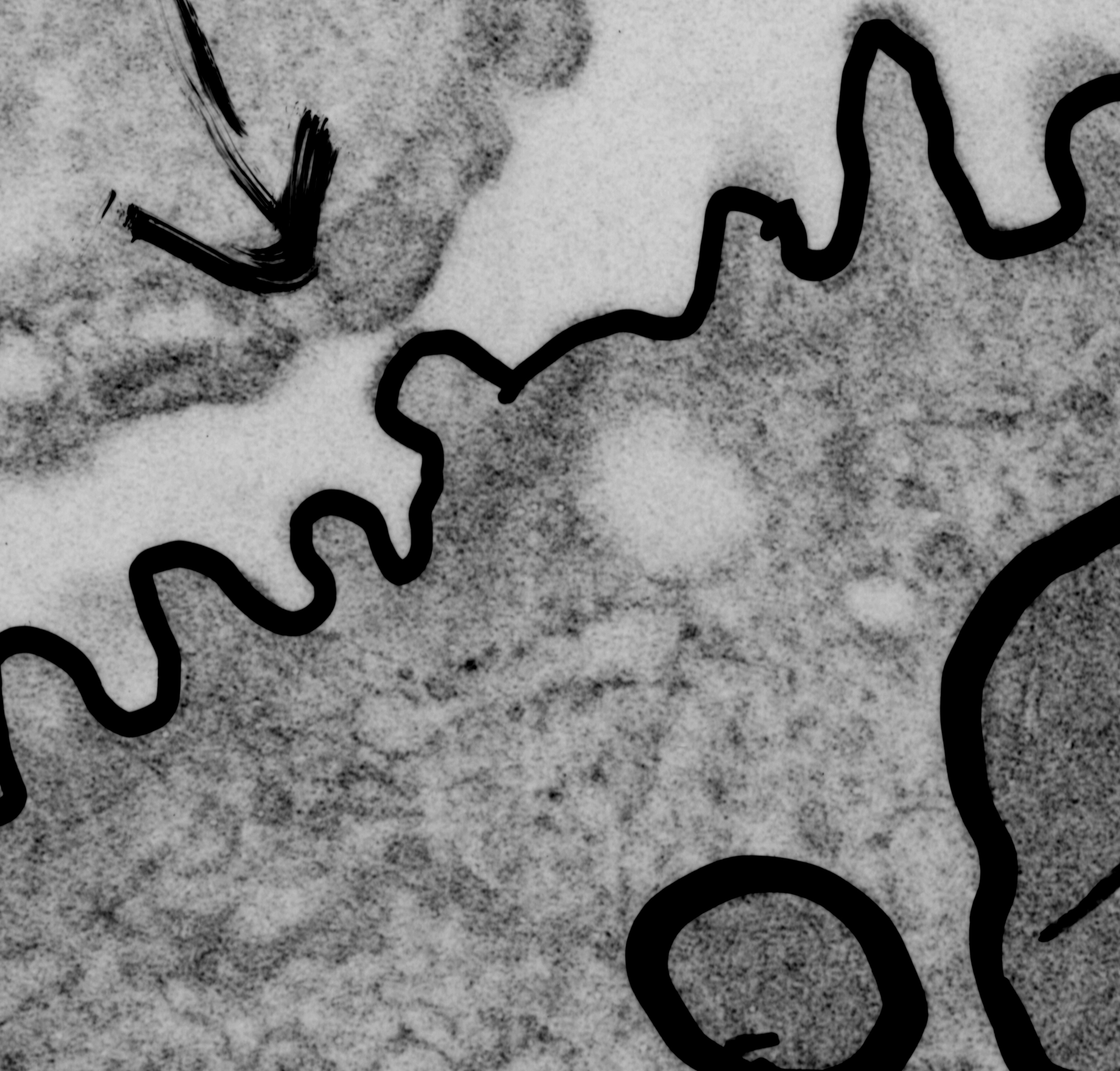
Lung tissue harvested from a sham treated control from another investigators experiment (thereby saving time – money – animals) here is a piece of a type II cell viewed with the electron microscope. It has my markings (for morphometry, back in 1982) and a marker arrow pointing to an area of RER which was the closest thing i could find to an organized protein in the RER (as a continuation of the search for species which may have SP-A organization similar to that found in guinea pig and dog and ferret. In cat, though the total number of electron micrographs I have saved is not that great (a dozen or so) therefore the absence of such a layered intracisternal protein is not to be assumed). The ribosomes studding the RER membrane provide an estimate of size (each being approximately 25 nm in diameter – a general number for all species and conditions…. (generalities are not always good for much, but an approximate size is OK here).