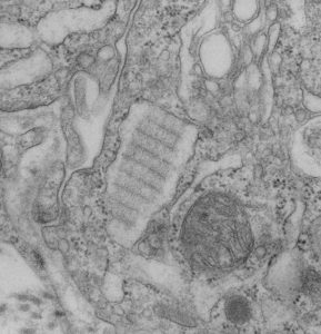
How awesome is this particular view of an intracisternal protein in a ferret type II alveolar cell. The bands are slightly tangential and so only the most electron dense banding is seen, however, the interesting thing here is that on the periphery there is not really any banding, but it looks as if the banding occurs centrally in the granule (aka within the profile of the RER). Also, in the upper left of this electron micrograph (in an adjacent type II cell) examine closely the profile of RER which actually can be perceived as ribosomes on the membrane surface and the trails of protein hanging off in the RER lumen. Really a classic text-book presentation in “real life” so to speak, though we all know that TEM captures only a “nanosecond in time”, and all proteins are “fixed” and so distorted. Nevertheless, this may be one of those opportune views.
The protein (which I think is SP-A) is central in the micrograph, the banding pattern is at about a 35 degree angle. Ribosomes are present on right and left borders for the most part, smooth ER is in the upper right corner and there is a tiny portion of a lamellar body off on the right side. A portion of intercellular space with a couple of plasmalemmal folds crosses from top center to bottom left. There are a few other profiles of RER without SP-A? banding as well.
