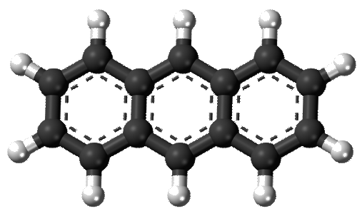
THis is anthracene – scanning tunneling microscopy by Bohringer and colleagues which has replicate images. It is perfectly obvious that a striaght line or rectangle and looking for peak height of grey scale is not going to give an accurate picture using ImageJ since sadly there is no option for drawing a line with a “node” for bending or adjusting the curvature of the line. Writing this into ImageJ would be impossible for the non-coder, but likely would be an easy task for others. If one wants to draw individual diagonal lines to compare with the LUT for a straight line however it is a fairly easy task. Because anthracene is so symmetrical it wold appear that not a lot of “remapping” is required.
In this particular case, cutting and centering the image (as proposed for curved molecules) would not create an acceptable dataset. Just taking a few minutes to examine this molecule with ImageJ makes it pretty clear that for large assymetrical molecules using the arc angle and cutting and centering might have some usefullness because of the speed with which the LUT analysis can be accomplished.
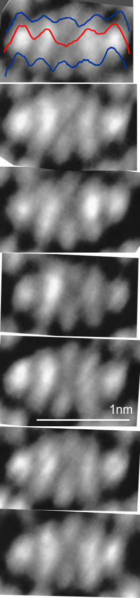
 These anthracene images (7 images below) appear very regular but there is a little bit of distortion angle to all of them, I but do not possess the expertise on STM or chemistry to say if this is artifact or not, but anthracene appears to be symmetrical. Red line is obviously LUT values straight across the molecule, upper and lower plots are 4 individual plots taken using ImageJ from edges of peripheral bright spots diagonally to the dark area in the center of the molecule. (i.e. four separate lines and four separate plots from upper right and left and lower right and left to center).
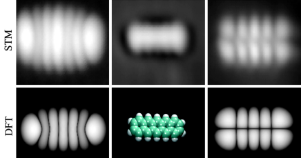
I added another diagram, because while the paper from which these images were copied says that there are a host of possible influences on the molecules being examined, and various changes in the appearance can take place. This image is what I believe is their control however, and i see no resemblance between their STM images (left and copied underneath on right) and the ball and stick diagrams overlying their STM images. ha. Most notable discrepancies in model and images occur at ends and in center.
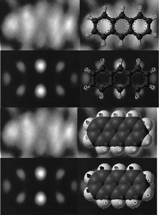 This image using STM (paper found here) is a more understandable to me, at least there is repetitive symmetry of the five rings between the ends. It ispentacene/2 ml NaCl/Cu.