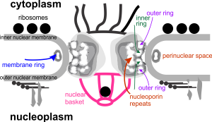
 This cropped portion of an electron micrograph of a nuclear pore was taken from the nucleus of a canine type Ii alveolar cell. It shows the filaments on the cytoplasmic side, and a bunch of ribosomes positioned just outside the nuclear pore, and two very distinct structures, about ribosome size that are either coming or going through the nuclear pore. It made me laugh, as it almost seemed as if they were going or coming in a “pin ball” fashion, being shot through the pore directly in the center. Do you see both of them? one is almost dead center of the micrograph, abd just elow the nuclear pore, and one is almost directly below that.
This electron microscopic image prompted me to try to figure out what is known about the nuclear pore complex in general, and every diagram I found in the literature was just a little different, even that found on wikipedia had some elements that I could not assemble over top the micrograph (which is basically what i wanted to do), so the diagram which accompanies this fine structural view (ultrastructure) is way off… I would love to find the time to put together a better diagram.
