Category Archives: Nucleoar organization

nucleolar-organization, RNA translation transcription electron micrographs

Size and location of nucleolar bodies

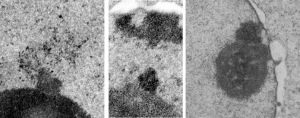
It is remarkable that these little segregated areas of nucleolar constituents look so consistently like “pop ups” that have a predetermined location, density, relative size, and texture.  I don’t believe that it is known what these early blips on the surface of the nucleolus are, (not including the blips that have the substructure notable of cajal bodies) though these may be precursors or similar structures. The banding (linear beads on a string appearance as if wrapped in a ball) in these bodies (by reason of tissue preparation or observer bias –shown in these three micrographs) would not qualify as cajal bodies. So what are they.  The bodies I am speaking of are all pointing upward like little volcanos (especially on the left)  with elements coming or going… whichever haha. Two images on right are slightly more electron dense than the adjacent nucleolar elements.

These images are from scattered micrographs, though two are from 14CoS hepatocytes which had never had or been withdrawn from NTBC.

Chocolate donut and sprinkles: nucleolar ultrastructure

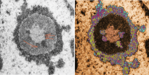
Shapes from biology (especially microscopy) sometimes are reminiscent of the most mundane objects in our daily lives. This nucleolus has reminded me of a donut with sprinkles for several decades.  In searching the nucleolus for clues to the connection between the sizes of apparent granule-fiber, beads on a string, things to what has been reported in the molecular structures online, I used this particular nucleolus (from a baboon that had been given perfluorophenanthrene emulsion (probably IV)) and some tissues biopsied. This is lung. In other views, this particular cell (not clearly identifiable but likely an alveolar macrophage) contains PFC droplets.

The “sprinkles” for this donut represent the dense fibrillar component (entities measured on a post from yesterday; the cake part of the donut is represented by the circular structure in the center of the side-by-side identical micrographs, which is the granular component. NB, the granular component on the left is light grey and round with the fibrillar center being the center of the donut.  It was noted in some of these nucleolar structures that there is such definite banding and that the lumps on a string is a very good casual description of the organization of RNP along a template and the measurements are pretty good.  As a reference point the diameter of a ribosome is about27 nm and the space between ribosomes in this particular cell is around 58 (or 2x the diameter of a ribosome). The diameter of the beads in the granular portion is 21nm, with a distance separating them of from adjacent structures is quite large and prominent, around 42nm (twice the diameter of the granule and though smaller in overall size than the ribosome and its space) is quite similar to it. The granules on a string in the dense fibrillar portion of the nucleolus is close to 30nm, slightly bigger than the average ribosome, and the space around these larger granules is the smallest among the three, at 38nm.

A second organization to the granular portion is seen in the parallel alignment and even spaceing of the granue. Orange lines lie over the very obvious linear pattern in the granular center of the nucleolus. A similar linear alignment is found in the dense fibrillar portion which becomes very very obvious in cajal bodies (not seen in this micrograph).

electron micrograph of the nucleolus of a baboon lung cellThis image would be a fun cover submission…  maybe someday i will make up a panel with three or four TEM look alikes. I know i have a valentine, and a ghost and a donut.

Granule sizes and spaces in the nucleolus

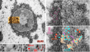
Epithelial cell in the lung of a baboon (probably a type II alveolar cell) showed a ring nucleolus with a single fibrillar center and concentric granular and dense fibrillar components. I took the time to measure (relative to a series of ribosomes measured in the adjacent cytoplasm – estimated at 27nm in diameter) the size of the granules in the dense fibrillar portion and in the granular portion of this nucleolus.  The image on the left shows the whole nucleolus, the three divisions (there are probably more) of the nucleolus are prominent. From that image of the nucleolus, the orange area is enlarged twice on the right to highlight distance between granules of the dense fibrillar component and the granular component. The small redish box is what was used to estimate the size of those granules by measuring ribosomes therin (red dot) obtained from a portion of adjacent cytoplasm from the same image.
The diagonal (and rearranged and measured as horizontal) lines are those between granules in the granular portion (white) and also the dense fibrillar portion (purple).  The spaces between the granules in the granular portion are marked out with a peach transparency and the blue circles represent the size of granules in that area.

Key: orange circles = size of the granules in the dense fibrillar portion at 30nm, with about 38nm distance between them; white lines and blue circles  21nm) represent the diameter of the granules in the granular portion and the distance between them (42nm).  For comparison, the ribosome (red dot, lower center-left is 27 nm, and the distance between ribosomes is about 58nm. The apparently-coiled strings (pink) are something that occurred regularly, kind of an arcing gently oval beaded appearance. 13279_94-32_baboon_lung_PFP (perfluorophenanthrene).

electron micrograph nucleolus monkey lung perfluorocarbon

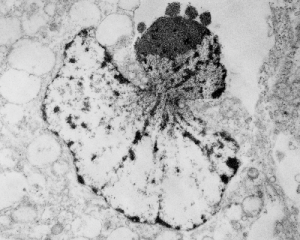
Asymmetrical apoptosis with four blips

I ask forbearance for my posting this. I have seen this asymmetrical type of apoptosis in some experiments that were done while investigating an anti-apoptotic gene. I was just doing the electron microscopy, others were doing the cell culture and knockdown experiments.  Typically apoptosis in culture presents with a round nucleus lots of large nucleoli and a bilateral (mostly) distribution of granular material along the inner nuclear membrane of the cell as it dismantles itself. The squeezing and pinching in this type of apoptotic cell was just really unusual and as is always the case on government grants, the reasons were never flushed out because of time and money constraints.unusual apoptosis in knockdown tissue culture experiment A549 cells I bet there is a really interesting reason for these, and this particular set of culture dishes got a scrambled version of an si67 probe and was supposed to be a control. It seems to me that part of the nucleus a just got extruded? I did count 8 nuclear pores (main criterion being an obvious set of cytoplasmic filaments off the cytoplasmic side of the pore.  Few mitochondria, many vesicles. One interesting thing about this apoptotic style is that the cytoplasm is not full of ribosomes as occurs in apoptosis in many cell types.

It is not something i could have made up (LOL) but my initial guess is that the four little ball-antennae are some remains of cajal bodies, and heretically i could suggest that the linear divisions radiating out into the “skirt” of whats left of the nucleus, likely has something important to do with each chromosomal territory (separations, walls, scaffolding, call it what you like).


Nucleolar ultrastructure: fibrillar center organization

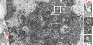
I was looking at this micrograph of a n hepatocyte with the fibrillar centers and dense thick fibrils in their midst, and the granular zone around the outside of this nucleolus. The latter being very punctate in appearance, just really obvious. I measured these once again at about 23 nm in diameter and noticed that around many of the fibrilar centers where was a basket, or ring, or radial distribution of these small punctate appearances.  OK so they are not really punctate, but can appear smeared like fibrils too, though the granular appearance and the nomenclature is well entrenched in the literature, they are just going to be strands not dots.  It could be that these radial dots around the fibrillar centers are bands, or windings at a given distance apart, kind of like fingers of a basket, or more aptly put, like a cage, with wires of 23 nm in diameter.

Electron micrograph here shows a portion of a nucleolus with three fibrillar centers cut out and enlarged (original sites are boxed in white), and a portion of the outer nuclear membrane with ribosomes (for measurement – one ribosome being about 27 nm in diameter, top right image with red dot and text). The cross section of the cage, or basket filaments around the fibrillar centers are shown as black dots.

Great humor: great truth

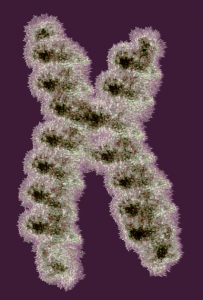
Reading about nuclear organization i ran onto this quote, and felt it was so great i just had to repost it. I personally continue to marvel at the naming of cellular structures, processes, and stuff, and the term “junk DNA” always rubbed me the wrong way, as does “non-coding DNA”.  I marveled to myself when reading those terms “what gives individuals the ego to believe they know enough to call anything “junk DNA”. I am showing a diagram of a chromosome (vector file with thousands of loops, and highlighted edges, iridescent graphic from a failed cover submission trial, a decade or more ago).

Well, here is someone who actually got that into a reputable journal… good for him — quote—from Thoru Pederson. Half a Century of “The Nuclear Matrix”, Molecular Biology of the Cell Vol. 11, 799 – 805, March 2000: “These kinds of ideas have been generally ignored because the noncoding DNA is so “uninteresting” as sequence (as if we were at present clever enough to be able to detect all “interesting” DNA text, which we certainly are not). At our present state of knowledge (ignorance) we can only view the noncoding DNA’s information content on the basis of what is absent [e.g., promoters, cap sites, splice sites, terminators, and poly(A) sites].”

Estimates of the size of some nucleolar components

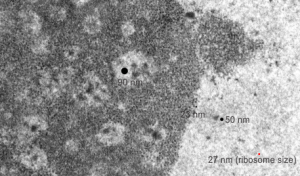
This micrograph, parts of which have been seen before, is an hepatocyte nucleus which is in the early stages of demise (untreated CoS14 ko without rescue). The nucleolus is large, and has two cajal type bodies (one seen here) and fibrillar centers with fairly large densities within (of approximately 90 nm diameter). I have given these sizes (relative to a ribosome size red dot approximately 27 nm (lower right hand side of micrograph labeled as such). The granules within the granular portion of the nucleolus (as well as the banding patterns found on cajal bodies) is something around 23 nm.  What I presume to be perichromatin granules look to be about 50 nm. The large dense bodies within the fibrilar centers are a little unusual.

Interchromatin granule cluster ?

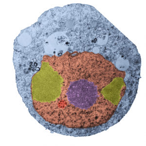
Nothing is black and white, except electron micrographs, and even then they are easy game for dodging, blending, blurring, mending, and pseudocoloring. Cytoplasm here is blue. In this case I have a cell from a culture of A549 cells in which C9orf82 was knocked down using si67.  This is pellet 4 from one of those studies. It produced this cell in which the granular component of the nucleolus was separated into at least two bodies (almost bilaterally symmetrically arranged) which rest at the inner nuclear membrane. Fibrillar centers are small, dense fibrillar component is still visible (albeit not distinctly) in the nucleolus (pseudocolored purple in the center of the cell).  Off to the side is what I could guess would be either degraded DNA (not my first choice as an answer (it is pseudocolored bile-yellow) or perhaps a fine granular component of the nucleolus in an apoptotic cell.  Part of an interchromatin granule (red, lower left) cluster (IGC) (sometimes called speckles, a name which is not really to my liking since the IGC has a boundary, a background texture that is also part of the structure).  So I would propose keeping two names, the “IGC” which is the greater boundary of that area and “nuclear speckles” as well, since the densities (as seen in this micrograph) can apparently be large.

If i am not off base here, the background of the whole IGC is red, but there are very clearly larger than typical speckles within the IGC, and the latter itself is quite small.  With immunohistochemistry the diffuse staining of IGC background proteins fluoresces with some of the antibodies to proteins like SC-37 in an area which can be 2 microns across easily.  I am looking for alternative proteins stained in very punctate regions of the IGC and hopefully they will be different and good markers for a separate ultrastructural components of the IGC. The point would be to localize to the densities with the IGC, some proteins for splicing, and accept that the diffuse staining could apply to SC-37, but not other proteins, mainly those in the variable size granules within the IGC.

We will see if there are data to back up the keeping of both names but assigning them to their obvious separate entities. BTW, there is pretty obvious bilateral symmetry to this apoptotic cell nucleus. and a radial symmetry to the dense bodies within the red colored IGC. And while i have no clue what the curley-Qs are… i bet they have something to do with the transfection. Just looking at this cell shows one tiny surviving mitochondrion, in this apoptotic cell, which is very close to the cytoplasmic filaments of a nuclear pore…also something which needs to be monitored during apoptosis.

Components of the interchromatin granule clusters are ??? and do they change during apoptosis

electron micrograph A549 cell knock down antiapoptotic geneWhile interchromatin granule clusters were thought to be the “the same structure” as nuclear speckles, i wonder if that opinion was justified at the time, I am not about to make that leap. I need to sort this out, since with plastic sections stained with toluidine blue, the interchromatin granules really were not “lucent or unstained” areas, but a light tan color, if i remember right, and with TEM they were actually more closely punctate than usual euchromatic nucleoplasm. So the connection needs to be further varified…and this might be why there are mixed notations and confusion and disparate explanations as to what the speckles and interchromatin granule morphologies identify. Also, what I have seen referred to as interchromatin granule clusters have two (at least) maybe three or four sizes of granular elements depending upon metabolic or functional state of the nucleus is (as in G,S and also stages of apoptosis, maybe necrosis as well.  (Of course all the (11 so far) listed processes of cell death and cellular-self destruction could be listed here but I just subscribe to the main ones, necrosis and apoptosis for now.) This cell has nucleolus dark blue, nucleus golden, cell cytoplasm greyscale,  interchromatin granule cluster of pink pink, and white box shows area enlarged in picture below. The larger (one quite large) and four or so smaller clusters can certainly point to some metabolic phenomenon which is not really occurring in a non-cultured-non-exposed cell.

The purpose here is to comment on the various sizes and shapes of such densities within the interchromatin granule clusters, and to examine transmission electron micrographs of so many cell-death projects to see whether concistent patterns interchromatin granule clusters have components change in size, position, density, and shape depending on which processes are occurring within the nucleus.

In particular, when A549 cells were examined after knockdown of an antiapoptotic gene (using si67), then the interchromatin granule clusters contained large aggregated densities (large in comparison to the smaller entities in untreated cells).  Here I have localized these in a single cell (transmission electron micrograph of the area bounded by the white box above), just to point out what is observed. Relevance is yet to be determined for these larger than usual areas of of conspicuous density within interchromatin granule clusters. The curved arrow points to bar-like organization of a filament or fibrilar area (sometimes this ribbon type linear organization is seen in cajal bodies) and the width of these bar-like fibrilar structures would be something around 30-40 nm, if compared to the size of a ribosome (red dot in the center of the text that says 500 nm).  The large structure (electron dense) beneath the interchromatin granule is the top part of the nucleolus seen in the image above. neg 18272 block 78932 A549 cell si67 knock down of antiapoptotic gene in vitro. electron micrograph A549 cell knock down antiapoptotic gene interchromatin granule cluster)