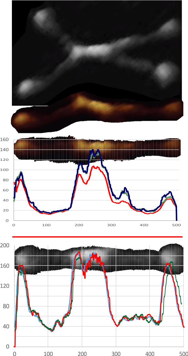
 Plots for the second dodecamer arm in an image publishes by Leth-Larsen et al,J Immunol 2005; which seems to provide similar counts of LUT peaks within the collagen-like domain of SP-D. I analyzed the first of two SP-D dodecamers in the AFM published images. One half arm with 1, then 2, 3 and 4… so all over the map. Plots were taken with 500×25, 500×17 and 500×1 pixel rectangles and superimposed upon each other. Two arms analyzed separately as on 500px length=100nm.
Counts by and (with out the LUT) were similar to peaks.