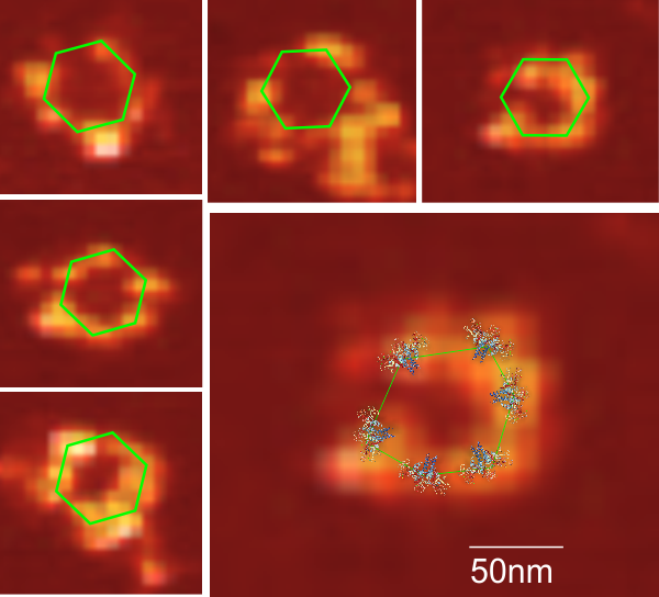
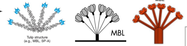
Nice atomic force microscopy of mannan binding lectin by Dong et al, though I could not use my method of measuring LUT plots on them. Nevertheless, in some of their images there was clearly hexagonal distribution and I have rearanged, marked and reposted some portions of their AFM image here and superimposed the carbohydrate recognition domain, at a slight angle (as would be projecting up from an N terminal (not shown) binding of six trimers. Many many (almost all) other researchers diagrams of show MBL as an oligomer with 6 trimers, not just three. The selected AFM images are “top down” the angle being 30o, the angle of the three diagrams is from the side. I don’t know why the difference here. (top image by Dong et al, bottom image is from google images no authors listed in this post but they are readily found online).