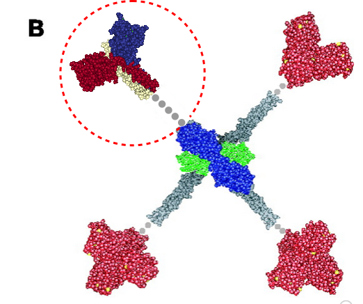
More confusion in the literature surrounds diagrams (in my opinion) than text. Here is a diagram which I am not quite sure how to interpret, however, this publication states that the grey dots represent an as-yet-undefined collagen-like domain in mannan binding lectin (protein) (MBP 1HUP – MBL2, and 1KWT) and I have read countless times and tried to determine its structure myself for both SP-A and SP-D and not found anything of high confidence. Yet the grey dots to infer “no yet determined” works for acknowledging that the molecular structure of the collagen like sequence in MBP (or the other C-type lectins) just hasn’t been worked out.. no that is not the problem, but it is that in my interpretation of this illustration, the grey dots are where the “coiled coil neck” is located NOT the collagen-like domain. So in the RCSB, 1KWt) entry, (shown against the original diagram published by Teillet et al, 2005, I have pasted the image from the RCSB (which includes the high confidence structure of the CRD and the coiled coil neck) against the CRD and the “grey dot collagen like domain” they drew. You can easily see my dilemma. My overlay is three color-for three chains in the trimer – of the CRD and neck, and grey dots for what I would have thought would be designated as the collagen-like domain, which then stops at the N terminus juncture (they show models of the serine proteases at the N terminus). The lower left and right and upper right arms of this cross-shaped mulecule have the dots (which they claim are collagen-like domains — where i think the coiled coil neck should have been drawn.
