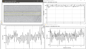
There were some really outstanding images of negatively stained tobacco mosaic virus in this publication. While reading the materials and methods it was like a throw-back ten years to staining grids for TEM. The disappointing part is not with their images but in my trying to use ImageJ to plot out the very obvious patterning, and the central “hole”, but this did not happen. Upper left is their TMV image (screen print), and three plots, one central line, lower left, one line at the edge of the structure, upper right, and one rectangular selection, lower right and also shown as the yellow-rectangle in the upper right image, in ImageJ.
Since the pattern is so obvious, i expected something much more informative in the plots.
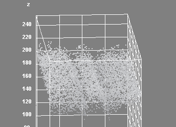
 Interactive surface plot in ImageJ shows up center much better.
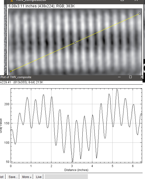
 The best plot is perhaps the angle line drawn over their composite TMV image.