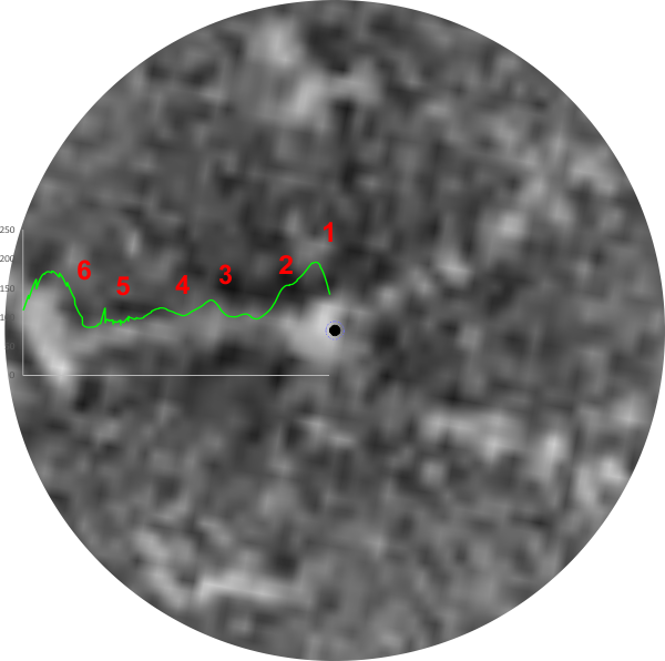
Of the images of SP-D whether trimers or dodecamers or multimers and whether prepared by different researchers by different methods, human SP-D appears to have 6 peaks along its trimer – first and last peaks being the Nterminus and CRD, second peak next to the former is the next highest and is possibly the glycosylation site occurring in the collagen-like sequence, peaks three and four are smaller and sometimes 5 which might be the neck domain is lateral to the former. All those are easily seen on AFM images, can be seen as well on rotary shadowed images. Here is a LUT plot from an image by Perino et al which is the third technique, that is negative staining. IT comes to mind that negative staining might be an interesting place to see these peaks, as the background noise, while greater than AFM – which smoothes over the peaks, minimizing them, is less than rotary shadowing, which has a background noise which is sufficient to mask some of the peaks. Thus the latter is probably more apt to show detail than either of the other methods.
So AFM, TEM of rotary shadowed SP-D molecules, and also negative staining show evidence of 5 (give or take) total greyscale peaks per trimer. What would have been nice is to have had a dark “hole” in the center (collection of Ntemini) of this multimer.
