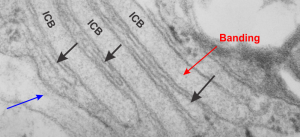
This electron micrograph shows three very long (a portion pictured here) lamellae of RER with a surfactant ?? protein, maybe surfactant protein A accumulation which shows a very very light area of banding – ICB) , and sandwiched inbetween these three are flattened cisternae (black arrows) which do not apparently contain the protein accumulations that is present above and below each. Whether these flattened cisternae without apparent protein are going to be sectioned down the block as thickened lamellae with protein content, is up for grabs, but likely (see lowest black arrow which points to a thickened part of the flat cisternae). This configuration is a little unusual. Other things to observe in this electron micrograph are: part of a lamellar body – upper right; tiny hint of the central band typical of what I think is overproduction of surfactant protein A – red arrow; basement membrane beneath the alveolar type II cell, and what is probably an endothelial cell – blue arrow;
