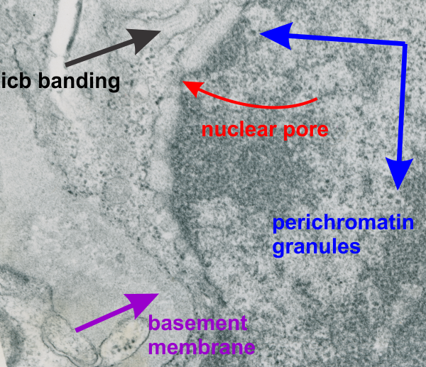
Nucleus, other organelles, nuclear pore, intracisternal protein accumulation, perichromatin granules, and part of the basement membrane are labeled in this electron micrograph of a type II cell from the lung of a guinea pig. Nucleus is on the right and a very thin band of cytoplasm is present, then there is the extracellular space (with basement membranes and parts of other cell processes). Basement membrane – purple arrow and label; red arrow and label – nuclear pore; black arrow and label – intracisternal protein which might be surfactant protein A (overproduced). Perichromatin granules (adjacent to heterochromatin) are shown by blue arrows and label.