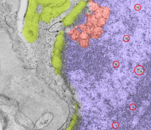
Looking at an electron micrograph of a guinea pig alveolar type Ii cell, (my guinea pig number is 301 – block 17082) archival material from the 1980s, I found this really interesting pattern in the nucleoplasm just above a nuclear pore. On either side of the outer nuclear membrane spanning the nuclear pore was the granule protein stuff that I have been working on, trying to place it possibly in the surfactant protein family, but being overproduced to the extent that it becomes its 18-mer, in mirror and/or vertically flipped position, times 2… producting the 100 nm banding that I see. But for this nucleus, just to the right of the nuclear pore marked with a two headed arrow pointing to the lime green intracisternal protein within the RER, there is a pattern that just “popped out” to me which is quite large (relative to the size of a ribosome…. and NOT to be confused with the hexagons in a tangential view of a granule but in the NUCLEUS proper) and sort of hexagonal in shape (which is what one expects when pressing together spherical bodies which I pseudocolored a salmon peach. Red circles surround structures that look like perichromatin granules (I am sure I could have found more).
