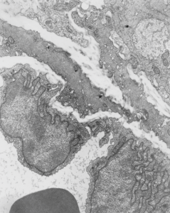
Feb 19 1975 under the auspices of the Children’s Hospital Mental Retardation Research Foundation (no longer in existence) house in what was called the IDR building (Institute for Developmental Research – also no longer in existence) with Leland C. Clark, Jr, I was in charge of looking at histological changes from his experimental procedures using perfluorocarbons: as 1) possible components of artificial (or synthetic) blood, 2) for contrast agents in lung, 3) and for lung lavage and aeration fluids. From the collection of animals and micrographs, here is a portion of a red blood cell, taken from an awake swiss albino mouse who had breathed E2 for 3 hours, and allowed to recover (apparently uneventful) for 1/2 hour.
I am totally not sure if anyone in the world will care about these data, but could not in good conscience allow some of the images to go into the recycling bin without recording them for posterity. Given long life, I will at some point assemble them into a manuscript, to submit “where” ha ha. Just relating this to the presumptive SP-A cisternal bodies (granules) that I am working on, none seen in this particular mouse (11 micrographs with type II cells at sufficient magnification to assess the RER and the perinuclear membranes.
Neg 1401, Block 4960, mouse 18.63 g, mag at scope=8,000 x, enlarg=2.83x, Karnovsky’s fixative instilled in trachea, osmium post fix, EPON 812 embedment. Original description of section with LM = general increase in cytoplasmic lucency, no plately plugs, blood vessels (per this photograph) were mostly open. Not seen in this micrograph but present in others to come, surfactant hypophase was abundant, the cytoplasm to nucleus ratio seemed increased, no E2 inclusions were found in alveolar macrophages. Red cell within blood vessel at bottom, endothelial nuclei diagonally across middle of micrograph. Plenty of “transforming fibrils” as Dr. Lowe and Dr. Charles Basom used to call them are seen in the interstitium. No alveolar type II cells in this image.
