Mouse lung, here is one of a few of the electron micrographs taken from lung tissue after various numbers of hours of liquid breathing, in this case the fluorocarbon is PP5, the duration of time was1 hr, with a 1/2 hour recovery. The negative is my number 1489, block number 5473, just in case there is interest in this image I can provide a higher resolution image or some additional backup. I recall that some of the perfluorochemicals used in the liquid breathing experiments seemed to cause a clumping of platelets in the very small vessels in the peripheral lung. I was struck by this early on, and I did not occur with all PFC, though I will need to confirm this. This particular mouse (4-7-1975) lung showed inclusions of PFC, and displayed some of the vascular characteristics that I have seen with liquid breathing experiments before: specifically, the accumulation of platelet aggregates in the small vessel lumena.
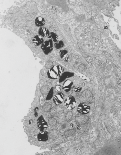
This cropped area from a micrograph just focuses however on the two alveolar type II cells which show two anatomical characteristis which I think might be related to liquid breathing of highly oxygenated fluorocarbons, 1) the lamellar bodies are very compact (maybe even categorized as slightly “immature” 2) the mitochondria look like they are on “speed” ha ha. wide mitochondrial matrix areas and cristae pressed to the sides and possibly reduced in surface area. Those two ideas are reserved for a later date.
This micrograph is a little out of focus, and is highly magnified from the original, quality is admittedly not great, but it demonstrates the point. The initial purpose was to look for the incidence and morphology of intracisternal RER granues in alveolar type II cells of mouse: (none found in these randomly saved micrographs).