All posts by thankusc

8 dodecamers of SP-D

Two sets of measurements have been added to the dataset for hexamer (that would be the CRD to CRD measurement of two trimers with N terms meeting in the center of the dodecamer) (Numbers of the molecules are my assignments  (out of about 90 different images) and number 127, and figure 4A,  are both images of the same molecule, but found in separate figures from Arroyo et al). Different image processing apps have been applied to strengthen the signal from the peaks from all images, and each image then was subjected to signal processing peak findings (5 different apps and settings which are now used for on all plots of images).

The data for hexamer width have really changed from previous analyses, i dont think more will be useful in determining the hexamer width.  I will still do this (just to find outliers and potential mistakes) in the upcoming images that I analyze.

Early data is on this blog….  you can check if you like.

Feb 23 data with 8 molecules processed in almost 900 different plots is summarized below.  The diameter and the length of the arms is calculated separately (in nm, relying on the nm bar markers in each image used).  THe total number of times the hexamers are measured is less than the total number of times the trimer widths are plotted since the same image is used for the signal processing (thus repeating it, while it makes very little difference in the statistics) has not been done here.

Verge of a dream: The same for us

I did not know
or expect
more than I
should
when returning
was what was said.

As promise was
not a word
You used or
maybe not
for me. And could
only guess
for anyone else.

I did not know
if coming back
was to me.
I’ve always felt
you knew
too much to
talk about truth.

I did not know
and would not
ask or
want an answer
but remembered
is too faint a word
for what I felt as
now wondering if
The embraces
were more or
less the same
for us.

RLB 02-10-2023

Zippie: life with mastocytosis and an aggressive mast cell sarcoma

Common in dogs, apparently rare in humans, my dog Zippie (around 13.5 years old) lived with this condition and not until after I put her to sleep (having a really aggressive mast cell sarcoma did I understand all those behavioral (odd, and troublesome) issues that she delt so well with.

In general it is thought that tumors composed of mast cells, which are a type of blood cell. Mast cells (with other white cells) are involved in innate immunity, and influence the immune (inflammatory) response of the body to foreign proteins (allergens). ,Increased mast cells (mastocytosis) affects blood forming cells of the body, that includes spleen, liver, gastrointestinal tract, and bone marrow, but also can appear as dermal tumors just under the epidermis, as mast cell sarcomas.

Quotes from the internet:
“These tumors share a universally bad prognosis with survival times of less than 4 months. They behave aggressively and require more aggressive therapy. These tumors overall showed a median survival time of greater than 2 years but there were some tumors that behaved aggressively despite their low grade.

Can dogs recover from mast cell tumor?
Well-differentiated mast cell tumors in the skin often can be treated successfully by aggressive surgery. High grade tumors are associated with aggressive tumor behavior, and most dogs die within 4 to 6 months after surgery as a result of metastasis and tumor degranulation.”

“Mastocytosis develops when mast cells increase in number and accumulate in tissues over a period of years. Mast cells are part of the immune system and are normally present in many body tissues, particularly the skin, lungs, and lining of the intestine. Mast cells produce histamine, a substance involved in inflammatory and allergic reactions and in the production of stomach acid. Because the number of mast cells increases, levels of histamine increase. Histamine can cause many symptoms, including digestive problems.”

“Cutaneous mastocytosis usually occurs in children. Occasionally, mast cells accumulate only as a single mass in the skin (mastocytoma), typically before age 6 months. More commonly, mast cells congregate in many areas of the skin, forming small reddish brown spots or bumps (called urticaria pigmentosa). Urticaria pigmentosa rarely progresses to systemic mastocytosis in children but may do so more often in adults.”
Systemic mastocytosis usually occurs in adults. Typically, mast cells accumulate in bone marrow (where blood cells are produced). Often, they also accumulate in the skin, stomach, intestine, liver, spleen, and lymph nodes. Organs may continue to function, with little disruption. But if many mast cells accumulate in the bone marrow, too few blood cells are produced, and serious blood disorders, such as leukemia, can develop. If many mast cells accumulate in organs, the organs malfunction. The resulting problems can be life threatening.”

In Zippies case, I think she had systemic disease (mastocytosis) and that seeded a mast cell tumor in her lower leg, and ultimately a large aggressive mast cell sarcoma on her outer thigh.
Quote – “affect dogs of any age but typically affect middle-aged to older dogs” (from Laura D Garrett). Clinically, heavy reliance is placed on the MI for behavior prediction (from Laura D Garrett). “The subjectivity and variability between pathologists bring into question the heavy reliance placed on the grade of an MCT to predict its behavior (from Laura D Garrett).

Qutoe “Mast cells and basophils as effectors of the allergic response” (http://what-when-how.com/acp-medicine/allergic-response-part-2/). If a release of granules from a mast cell takes less than 60 seconds, then a trigger can be seen almost immediately. Mature mast cells in peripheral tissues may reside there for many months, retaining antigen-specific IgE for periods that exceed the lifespan of IgE in the circulation. Mast cells are strategically distributed in tissues or at mucosal surfaces that interface with the external environment; they are also in proximity to blood vessels and nerves (see prior link).

Flushing is common. may develop because too much histamine is produced, stimulating secretion of excess stomach acid. Ulcers can cause stomach pain. Nausea, vomiting, and chronic diarrhea may also occur.

First line of this commentary on mast cell disease is going to include my sadness, and also a sincere amusement at the places grief can be trigggered: In the canned tuna aisle in the grocery store, and the relationship will become clear.

I found Zippie running lose near the tennis court behind North Avondale Montessori School. I remember seeing two little boys and they were intrigued with the dog and I lent them my leash (from a current rescue Zodie who was at the tennis court with me) to take the dog and ask their mother if they could keep her. (obvious result was the mom said no, and they brought the puppy back to me). We went to the vet for shots, and exam, and her approximate age was 3 months, and that date was Sept 16, 2009. Then we just began the process of getting her adjusted to the dog I already had.

There were not really any health issues with Zippie that would have suggested that she had a mast cell issue when she was young, though there were obvious signs that she had an immune system that didnt function very well: ear infections, diarrhea, urinary tract infections. I took the number of incidences as just a little worse than normal, not really that troublesome.
Her energy level and interest in walking and playing was pretty normal.

Middle age began to show serious food allergies, and symptoms of mastocytosis, and this was before any subcuteous tumors were present.

Oakley Sq Annimal Hospital removed a subcutaneous lump from the front of Zipper’s leg on Aug 3, 2020. This tumor (skin and tumor were movable on the underlying tissue, and dense, and waiting to remove the tumor would have resulted in not being able to find skin to close the incision. I dont know whether an adequate margin around this tumor was possible. This is just over the predicted survival time after a mast cell tumor has been removed, she lived from that surgery until Feb 3, 2023, so just over two years. The last two weeks saw tremendously rapid tumor grown and increasing evidence of mast cell degranulation (in terms of shivering, panting behavior).

References:
Thank you to the authors of the following articles

Verge of a dream: It could be about anyone

A lot we know and
So much we don’t
as it could be
about anyone, true.
The life was not
bad,
Mine, yours,
with laughing
at times, it missed
by inches, or days
Or other souls
being worse.
For very last
acts, after all
have been done
and
whether in words
or ideas
they will not
compare or preach,
or judge. Then
I won’t think
Of you with
Someone else
Skilled and knowing
Painful In dreams.
In denim, you
Move toward the
Water. Glimpses of
A suit in two
Pieces,
When a
plaid blouse
Slips from
the shoulder
and the sandy
wash returns to
the ocean.

RLB 02-6-2023

Verge of a Dream: Unveiled

Some things that don’t
And some that matter
Your white sweater
Clinging, an oriental
Blanc de blanc
To you until unveiled
Or until the end
Of suffering by,
There is no way
To know how,
Among that which
Does not matter, by
His own hand.

There are times
And some without time.

Not watching
A newsreel
Because in watching,
Watching is something
That will not and wants
Not change.
Except, wanting parents,
That change, that now
They are gone.

Give me a gift.
Hold me when
You worry
Give me two gifts
Love me without
Feeling sorry.
I won’t ask for three
Though so many
There were to give.

RLB 01/30/2023

and an edit


Some things that don’t
And some that matter
Your white sweater
Clinging, a
Blanc de chine
To you until unveiled
Or until the end
Of suffering by,
There is no way
To know,
Among that which
Does not matter, by
His own hand, how.
There are times
And some without time.
Not watching
A newsreel
Because in watching,
Watching is something
That will not and wants
not change.
Except, wanting parents,
Only
that now
They are gone.
Give me a gift.
Hold me when
You worry
Give me two gifts
Love me without
Feeling sorry.
I won’t ask for three
Though so many
There were
Left to give.

6 dodecamers, 368 trimer plots, peak width, height, valley plots

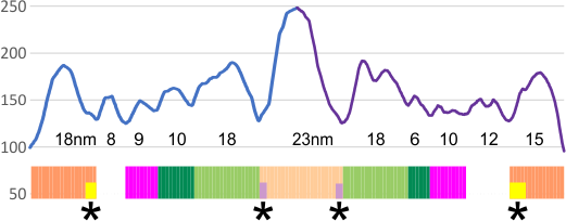
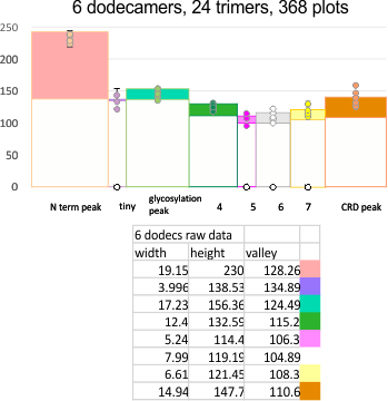
EDIT: the plot shown below is what I think describes the data. It has the mean peak height, peak width and peak valley from 6 molecules. No smoothing or blur or anything else, just the numbers. It might be as useful as anything that some algorithm can invent.  Peak width is x, mean for the six molecules (the number of peaks per trimer and hexamer was determined by signal and image processing data early on) (15) and per trimer ( 8) respectively.

Number of peaks from each program depended upon various parameters, lag, threshold, influence, smoothing, and many I dont understand, but the separation of the signal and image processing graphs into the 8 peaks (color coded) was was performed by me, which, in my humble opinion, is just as good, if not more “learned” than any AI app.

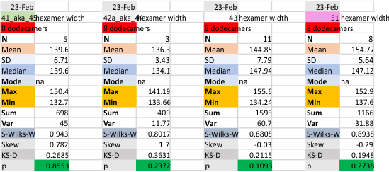
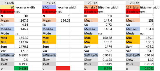
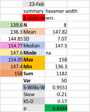
The separation of each hexamer (or trimer) into peaks is reasonably consistent in terms of peak width, height and valleys.  THus I have means for all tracings, means for individual molecules, plus SD for widths, heights, and valleys, all of which can be given in table form shown below.

Means of all plots, and individual trimers dodecamers can be shown in the style of graph below, with SD of each parameter. The top image here is just a quick graphic of what that kind of plot would look like,  and are close to the actual numbers below but not exact, as this is a draft.  Peak width is in nm, peak height and valley are in grayscale 0-255.

Anyway, it is the format that I will use for collecting data on the remainder of the SP-D molecules.

Other options that I worked with for plots are below –

I have extensively looked for peak width, valley, height plot apps and cannot find one that works for me…this doesn’t mean they dont exist, but i have not made the effort to get on the chats in scipy and octave to find them.  The basic set of numbers is super simple, there is the possibility that i have not collected them in a way that is useful for making automatic plots.  They are perfectly useful for constructing a plot using a graphics program however.

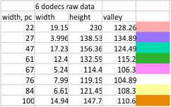
Height and valley values are in grayscale (0-255), width (has variable measures (pixels, inches, cm and is not consistent) is changed to percent (left column).

Basic numbers for the width, peak height, and peak valley (data from the valley closest to the N term side) are here. Help is certainly welcome. Data below was accumulated from image and signal processing one hundreds of plots of the AFM images of surfactant protein D. Previously it was determined that the mean number of peaks per hexamer was 15, that means in counting the trimer peaks, the N center peak gets counted once for each trimer, but also, only once per hexamer, thus the number of peaks per bilaterally symmetrical  hexamer which is comprised of two trimers (but the N term blends into  a single very bright peak) is an odd number.

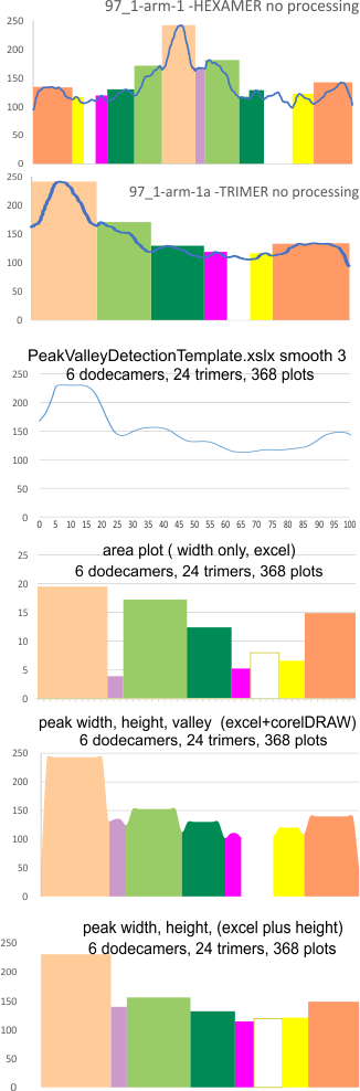
Example of a real plot of a SP-D molecule as a hexamer is top… below that is the same plot trimmed keeping the N term peak (light orange) as a whole, not dividing it into have – part for each trimer.

Rhe trimer plots  below assembeled in various ways with various problems and various programs (but mainly excel and corelDRAW). From top to bottom, beginning with the pinkish peach color N term composite peak (peak1); tiny peak, purple (peak 2);blue-green, glycosylation peak (peak 3);  darker green, peak 4; narrow peak 5, pink; unknown peaks, white, coiled coil neck domain yellow, seen intermittently, and not seen when it is likely to be behind the yellow, and last peak equals CRD peak.

I dont think this is rocket science, i just need to find the right program and certainly the data are consistent with the numbers in each case, just not “pretty plots”. In the case of Peak Valley Detection Template xslx, there was no value between 1 and the next highest smoothing function (3) that would do a better job of keeping the peaks but smoothing the corners.   So this is a “taste” thing, not important.

So the issue becomes how better to collect the data.

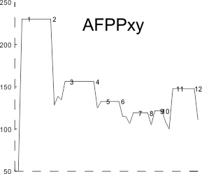
Here is a cute thing — actually not so funny… the summary plot .csv file plugged into octave, I was hoping to smooth the plot, and here i find that the corners of the line plot count as extra peaks….  Clearly, this plot has 8 peaks… not 12, and it didn’t bother counting the “tiny peak” (purple in above plots) or peak 5 (pink in above plots).

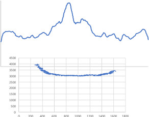
I found a link to an online converter of svg to matrix called Coordinator.  I put in an actual plot (see top image below) and used this open source app to create a plot.  It was not exactly what I had thought  (smilie face below)–  as i had been thinking for a couple years that I would really like to use the graphics flexibility of corelDRAW on the excel plots, then convert the vector graphics back into  a matrix…. didnt work that well the first time…???

Just for comparison with the plots of this same molecule, several years ago before signal processing was in the picture, here are the number of peaks per hexamer (11), and the additional 4 peaks, not present 100, or even 60% of the time, are four peaks (two pairs) which show up consistently enough to be considered something to work out.

Parietal cell morphometry – dozens of KOs

These data are all performed on mice (KOs and treated) from the laboratory that was run by Dr. Gary Shull and his students and postdocs. I saved these plots (which were intended to be submitted as a manuscript back in 2007, when I retired) and am just now sorting through the data from those years in microscopy.  I did morphometric measurements on many of the mice (listed below) which were published, but gathered together those into a composit kind of analysis — not a publication, just a view into what could have been published had time permitted. I dont think at this point anyone will try to rebuild this, but the information as a “glimpse” into changes in the basolateral (i dont like that term, it is clearly a lumping together of two very distinct membrane domains, nevertheless it is in common usage), apical membranes, tubulovesicular, and other structures into a quantitative assessment over many different genetically modified, or treated mouse parietal cell datasets.

The graphs in the pdf represent hundreds and hundreds of hours just for the morphonetry, let alone the animal experiments, the creation of the animals.

The plots in this pdf (some shown below just so you know what is in the pdf) are low quality prints that I used just for reference. The actual data can be recovered with great effort but if someone wants the numbers, i will try to retrieve the original datasets. A pdf of all the graphs is HERE parietal_cell_morphometry_many_KO-mice1

Selected screen prints below”