Category Archives: Ultimate order, the cell

The beauty and order of life is astounding.

Great research on desmosomes, but

All you guys out there that are doing great research on the molecular structure of the proteins comprising the desmosome, BUT, there is a huge disconnect in what is visible with routine electron microscopy, and the diagrams that are being produced.  Just for starters, if I (not being an engineer but just a craftsperson) were going to build a rigid structure binding two adjacent objects i would NOT make it unidirectional.  That is I would not orient all the layers of the molecules in parallel.  I would do what is commonly done in ply wood, that is to plait the structure with perpendicular, parallel, perpendicular, parallel layers.

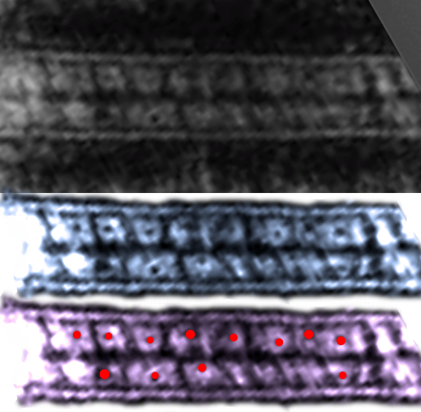
The way i see it in transmission microscopy, the desmosome has a set of proteins perpendicular to (and intersecting) the plasmalemma (desmocollins and desmogleins), then a row of proteins which act like lock washers or lock washers (plakoglobin, plakophilin and others I don’t even know about) that are pretty much perpendicular to the desmocollins and the desmogleins, and then another set of proteins, represented by desmoplakin, that becomes a row perpendicular to the to those “armadillo” proteins, and finally, the connection of desmoplakin with the intermediate (or other) filaments again perpendicular.  So there is a layering of different proteins, designed to accept all kinds of stress, and pulling, and shifting.

I have yet to find a diagram, out of literally thousands, which conveys this idea.

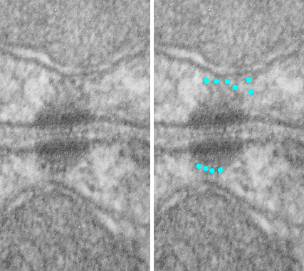
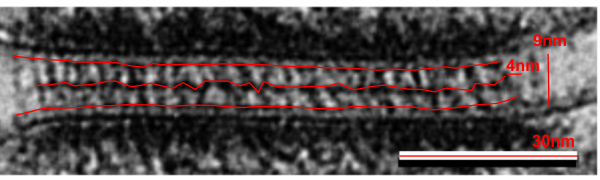
What’s more…. the fixation on the desmocollin desmoglein partnership creating the central dense line of the desmosome as being like a W does not fit the transmission microscopy either.  It would more likely be an S, again, rarely mentioned, except one pretty close approximation to what is seen (also an S and an Lambda shape).

A really disturbing depiction is the continued showing of intermediate filaments coming into the desmoplakin with a hairpin turn and back out again….. that just is not seen in TEM.

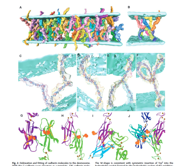
top diagram is what i have readily found in the literature…. mostly parallel depiction of the proteins which comprising the desmosome.  (black and colored vertical stripes) — which I think misses the point of the layering.  Most left hand bars are what I would predict, everything from the adjacent diagram to the right imply parallel arrangement of the proteins, including the intermediate filaments.

So this to me is more structurally sound, and we all know nature is pretty good at making changes to improve “soundness” LOL. So I would opt for this interepretation below. an explanation: plasmalemma (three layers blue and white); central band within the plasmalemma would be what is seen with TEM, i.e. the dense central line (dotted here) made by the association of desmocollins and desmogleins from each adjacent cell and this happens in the extracellular space; the armadillo repeat proteins, as many diagrams put (but not this one) are aligned more or less vertically along with the intracellular domains of desmocollins and desmogleins, but here I have emphasized the “lock washer, or washer” function holding two parts of this cellular rivet in place.  THen the desmoplakin molecules which in turn align perpendicularly to the area of the outer dense plaque proteins…. and finally again perpendicular to the desmoplakin… the intermediate filaments (green =  direction of orientation.

Mechanotransducer (the desmosome)

Mechanotransduction is known to be an important biological process rendering tissues both resilient and responsive towards their mechanical environment” says the first line of the introduction of this article.”–   mechanotransuction: definition:(biology) Any cell, etc. that generates a measurable response to mechanical stimulation–so this definition includes many many organelles, including the desmosome.

It is fun to think about the desmosomal – mitochondrial tether as the energy supply and the

Adherens junctions and desmosomes

Narrowing of the intercellular space = about 5nm where the cadherins couple

I am not sure why the intercellular space is recorded by What When How as different dimensions (beside the adherens junctions 25nm vs 20nm, the height of the adherens junction itself) and of the intercellular space beside the cardiac desmosome as 35nm and the height of the desmosomal intercellular space as 20-25nm. It seems to me that if one is comparing intercellular space heights, that one really needs to set some parameters and get some comparisons. This could be a massive job, as there are so many variables, not to mention fixation parameters, tangential sections, membrane proteins, cell types and where on the plasmalemma one is going to attempt to measure.
Just in four desmosomes (between hepatocytes from syrian hamster) all fixed the same, similar ages and reasonably good section orientation the following is clear.
1. the intercellular space is pretty variable
2. desmosomes are going to have a slightly smaller intercellular dimension than adjacent intercellular space
3. the center dense line extends beyond the outer desomosomal plaque proteins
4. there is a density in the plasmalemmae as an annulus or ring around the desmosome but intercellular space is wide
5. the separation of leaflets of the desomosomal plasmalemma is just a little more distinct than distant plasmalemma
6. the outer leaflet of the plasmalemma at the desomosoe seems to be quite rigid

See below, four examples and the relative wide range of reduction in intercellular space. Syrian hamster — routine electron microscopy, red dots intercellular space remote from the desomosome, blue dots height of intercellular space at the desmosome. Two of the images – right top and bottom – have double mitochondrial tethers. Left side images have a single mitochondrial tether. Right top and bottom have cross and longitudinal sections of intermediate filaments, respectively adjacent to the mitochondria. In both cases the outer mitochondrial membrane also has a rigid look.

Easter bunny mitochondria

Please forgive, LOL, as this little mitochondrion which is from guinea pig liver, just was staring out from the page. You might notice that there are some (blue) regular protein molecules near the bottom right and left.  These are likely to be ATPsynthase all aligned in perfect order — see previous post with similar structures from guinea pig hepatocyte mitochondria.

Bridge or intermediate filaments?

When I first looked at this micrograph i thought perhaps the electron density under the mitochondrion (here above two desmosomes, tangentially cut and off center, was a bundle of intermediate filaments. That would be interesting since i don’t know if filament bundles likely composed of keratins 5 and 14, could make this tethering bridge between desmoplakin –part of the desmosome in complex with two remote points on the mitochonrdion.

Turns out that It looks more like a little invagination along part of the plasmalemma of the mitcohondrion, probably representing outer mitochondrial membrane. Red dit=ribosome@27nm, blue curley bracket=bridge, red lines, intermediate filaments bottom red lines, underscoring position of thedesmosomes.

electron microscopy mitochondrion

Outer mitochondrial membrane tethered to intermediate filaments

There is a reasonably prominent increase in density (and rigidity) of the inner and outer mitochondrial membranes where they are tethered to intermediate filaments tethered again with desmoplakin of the desmosome complex.  It occurs for the stretch which runs parallel to the desmosome and removed from it by about 140nm in height (as measured on these two micrographs – not ideal cross sections of desmosomes, so approximate dimensions).

Someone out there researching mitochondria membrane proteins probably knows which proteins these would be.  Top electron micrograph shows two separate mitochondria (tethered  — albeit tangentially) to the desomosomal complex showing intermediate filaments parallel to the plane of the plasmalemma in both instances. Red dot, ribosome=@27nm, red lines=intermediate filaments, blue curley brackets=inner mitochondrial membrane with increased density, green curley brackets=outer mitochondrial membrane with increased density at points of tethering. Dotted line in photo bottom left is where the outer mitochondrial membrane likely travels.


Desmosomal mitochondrial tethers

It makes a lot of sense that there are mirror images between two cells  — that is in terms of desmosomal / mitochondrial tethering. I have seen as many as six desmosomes tethered to two mitochondria (in juxtaposed cells). It seems that if the mitochondria supply the energy and the Ca+ regulation (for unzippering the desmocollin and desmoglein) and signals for dissolution and/or construction of desomsomes that one mitochondrion could do the job for three or more desmosomes.  I can envision the mitochondria alligned up along the lateral plasmalemma like soldiers standing at attention ready to build or destroy…

    1. seems pretty certain that transmembrane  portions of desmocollin and desmogleins affect the trilaminar architecture of the lateral plasmalemma, clearly seen on electron micrographs — they stiffen it….and widen it.
    2. There is an also an effect on the lateral plasmalemma in the annulus of the desmosome, appearing to be slightly more dense than plasmalemma further away from the desmosome, and also with slightly wider extracellular space (than non adhering areas) from outer lamina of the plasmalemma of each adjacent cells.
    3. Seems pretty likely too that most of the diagrams i have found in publications on the topic which bring intermediate filaments to the desmoplakin molecules at a parallel or nearly parallel position are just not accurate. I see lots and lots of perpendicular structures, that is desmoplakin perpendicular to IFs, rarely if ever one that looks like the desmoplakin and IFs are running the same direction. In fact, it doesn’t make sense architectural sense (in terms of adding resistance to pull and shear and separation to have them run the same direction. The long polymers (the IF) run parallel to the mitochondrion which really remains a fixed (seemingly) distance from the outer desmosomal plaque proteins…and plasmalemma. It looks likely that there are 4-6 IFs or more that run parallel to the plasmalemma (and outer membrane of the mitochondria)  not perpendicularly to it.  Just my thoughts (and what I see).
    4. Here is an electron micrograph which has cute little cross sections of IFs in the space between the mitochondria and the desmoplakin and outer desmosomal plaque proteins. (see the blue dots in micrograph on right, which I interpret to be IFs). Left hand image unretouched from scanned photo. (I could have added at least 5 more blue dots to the image on the right where blue dots are overlying cross sections of IFs.)

Man never made a rivet like this: double sided ductile suspension shear load lap rivet

haha…. you think i am joking: just contemplate the desmosome

1) velcro (desomogleins desmocollins) rivet proteins with central hooks in the intercellular space
2) lapping surfaces (trilaminar membrane from two adjacent cells) penetrated by the desmogleins and desmocollins
3) intracellular lock washers (the plakoglobin and plakophilin linking to the desmogleins and desmocollins after their transmembrane pass) reinforcing the rivet body
4) suspension flexibility perpendicular to the cell membranes (desmoplakins which attach to the plakoglobin and plakophilin lock washers) absorbing both shear and
5) intermediate filament (ropes for applying resistace and tension to the desmoplakins which lie parallel to the plasmalemma NOT hairpin like most diagrams put them)

Intercellular organization of desomosomal proteins

Looking further at the great publication by He, Cowan and Stokes in Science 2003, i just was surprised to see what looked like the most orderly set of dots in the intercellular space on either side of the central dense line (where the desmocollins and desmogleins are supposed to be hooking up in their “velcro-like” attachments. I did a screen print of their micrograph (top image which I did not manipulate)and pasted into photoshop to enhance the pattern with the burn tool (shown in the two bottom images) as an overlay of two separate layers of their original image.  The dots and grid are so obvious as to be almost “silly” and if you don’t see what i am talking about…. check out the red dots within the grids in the two images below their micrograph.  I have looked at hundreds of desmosomes…. now i have to go back and see whether any of my more opportune images show dots within the grid (which I have seen before.  Here the Y and alternating pattern of the intercellular grid is very very apparent.

It is still possible i think, to have the Y formation be the most prominent, and also repeated in a very orderly fashion (one difference between what i see and what the paper above suggests… when they call the desomosomal cadherins to be in “a knot”.  I don’t think disorder is part of this structure… but there rather there is an order which is just a little difficult to detect, owing to the very many angles (at a thickness of 90nm) to bisect a desmosomal spot and untying those possibilities (not to make a bad pun on their title) is not easy.  U havn’t yet found a publication that speaks to anything that would make these central densities.

I know there are no mitochondria in their micrgraphs… and that is ultimately what I am curious about, but just in case you havn’t figured it out yet…..  biology is totally amazing.

3D intercellular space of desmosomes

This is an awesome representation of the intercellular space of a desmosome (that I am not giving a classification to…. either coming, stable, or going since apparently the intercellular area undergoes ultrastructurally visible changes, namely the loss of the central dense periodicity, as they change states).  The paper by He, Cowin, Stokes appears in Science linked here. Two reasons that I love but dont like this diagram. 1) it depicts exactly what can be seen with traditional transmission electron microscopy of the intercellular area of desmosomes, but 2) it doesn’t seem to ring true that the organization since a “knot” is less orderly? in my mind, more “chaotic”, and I think the electron micrographs show the intercellular organization is way more orderly than the diagram suggests. 3) the number of molecules in the colored diagram (part A as they label it) is more than I seem to see in the sections with TEM.  The banding (i am guessing most agree to be desmocollins and desmogleins) is so regular, and the space between so lucent, that all the knot like other molecules are likely overrepresented.

I agree that in this particular view here there is only a tiny hint of any symmetry. I do like that the dimers that they have highlighted (one actually as a lambda, my favorite representation, and the one that fits the intercellular space-repeating order of a desmosome in many of my own electron micrographs best —  lambda shapes mirrored vertically and ofset by half). There is no way here to tell if something was influencing the desmosmal structure (e.g. the presence of a very close mitochondrion, maybe participating in the formation or destruction of the desmosome in their picture).  Lots of opportunity for that variability to be a part of a process influencing what is viewed as a chaotic knot which in fact was a highly organized intercellular component of the desmosome. I am linking their image to their article.

(it would indeed be fun to have access to that equipment, and knowledge of how to use it, and possess the specimens made to order,  poof, all at once and with extreme precision).

This publication also shows some TEM images of desmosomes which i did some measurements with: the distances between the densities at the outer plasmalemma of both cells, and the distance between the periodicities of the central dense line of the intercellular space. All distances were similar… mean of all was about 4nm center of density to center of density.  The intercellular width (from the outer plasmalemma to the adjacent cell was about 9nm. See lines from that TEM below (link to the manuscript is above).