It makes a lot of sense that there are mirror images between two cells — that is in terms of desmosomal / mitochondrial tethering. I have seen as many as six desmosomes tethered to two mitochondria (in juxtaposed cells). It seems that if the mitochondria supply the energy and the Ca+ regulation (for unzippering the desmocollin and desmoglein) and signals for dissolution and/or construction of desomsomes that one mitochondrion could do the job for three or more desmosomes. I can envision the mitochondria alligned up along the lateral plasmalemma like soldiers standing at attention ready to build or destroy…
-
- seems pretty certain that transmembrane portions of desmocollin and desmogleins affect the trilaminar architecture of the lateral plasmalemma, clearly seen on electron micrographs — they stiffen it….and widen it.
- There is an also an effect on the lateral plasmalemma in the annulus of the desmosome, appearing to be slightly more dense than plasmalemma further away from the desmosome, and also with slightly wider extracellular space (than non adhering areas) from outer lamina of the plasmalemma of each adjacent cells.
- Seems pretty likely too that most of the diagrams i have found in publications on the topic which bring intermediate filaments to the desmoplakin molecules at a parallel or nearly parallel position are just not accurate. I see lots and lots of perpendicular structures, that is desmoplakin perpendicular to IFs, rarely if ever one that looks like the desmoplakin and IFs are running the same direction. In fact, it doesn’t make sense architectural sense (in terms of adding resistance to pull and shear and separation to have them run the same direction. The long polymers (the IF) run parallel to the mitochondrion which really remains a fixed (seemingly) distance from the outer desmosomal plaque proteins…and plasmalemma. It looks likely that there are 4-6 IFs or more that run parallel to the plasmalemma (and outer membrane of the mitochondria) not perpendicularly to it. Just my thoughts (and what I see).
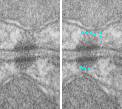
- Here is an electron micrograph which has cute little cross sections of IFs in the space between the mitochondria and the desmoplakin and outer desmosomal plaque proteins. (see the blue dots in micrograph on right, which I interpret to be IFs). Left hand image unretouched from scanned photo. (I could have added at least 5 more blue dots to the image on the right where blue dots are overlying cross sections of IFs.)