Toluidine blue stained thin plastic sections of parietal cells in the stomach of a mouse (experimental strain). Image processed, probably in photoshop. Would love to have a scarf with this print.
Category Archives: Science and art cover illustrations
SP-A and SP-D diagrams: these take the “cake”
Oh my goodness, how far from reality some illustrations, aka. scientific diagrams, are from the truth. I do not understand how individuals who are supposed to “know” a topic, a “protein” a function of something like surfactant can use a ridiculous diagram like this and expect to gain any credibility whatsoever. My gosh…. These diagrams have nothing at all to do with either SP-D or SP-A, they are fake news, provide conflicting understanding and do little to advance the knowledge of how proteins really look. So sorry, but there is absolutely no excuse for this lack of understanding and depth of falsity.
I wonder if i could establish a monthly award for the dumbest scientific diagram ever seen.

I wonder how this image came about?
It is interesting what individuals do for their microscopic images for publications. Looking over all the TEMs i can find for SP-D, SP-A, conglutinin, MBP, etc I find that some methods for presenting images are nice, they probably are NOT what I would have done. In 1993 when this manuscript went to press I don’t believe that very many individuals recognized the potential for photoshop to create “pretty” images.
Changing the contrast by dodging with fingers and spoons and holey circles was about the extent that an image could be manipulated in the wet dark room. Occasionally we huffed and puffed on a print to warm parts hoping for more development on one side or another but other than that, there wasn’t much one could do with a print to beautify it.
In this manuscript (Eur. J. Biochem. 215, 793-799 (1993)) the authors have performed (in my opinion) the interesting, unnecessary, somewhat deceptive task of putting three SP-D molecules (the dodecamers) into a straight line portion B of their figure. It didn’t take much “looking” to see that the same SP-D dodecamer is present in a field of their part A of that figure, and again with two partner dodecamers in part B of the same figure. While this doesn’t necessarily change their data, one was rotated slightly from the other, and just a tad different in magnification. The better approach might have been to put a white division line between the first, the second and the third SP-D dodecamer in panel B, rather than to fuzz the background together…. that was just on the verge of being deceitful, since it gives the impression that lots of routine dodecamers appear close together in their prep. Probably not the case. That said. Below is their figure part A with the single dodecamer, and part B where they have cut pasted and blended the shadow background. You judge.
It might be noted that journals right about this time built (or were supposed to have built) algorithms to detect manipulation of the pixels in publication images.
 And just in case you are skeptical that they are the same image, see the radial pixel pattern upon rotating half transparent images one from part A and one from part B just a few degrees (which doesn’t happen on non duplicate images (again, in my experience)).
![]()
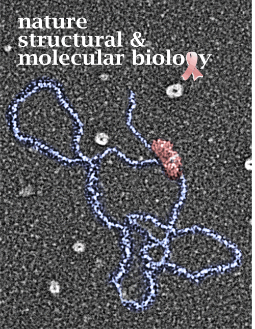
Cover submission – breast cancer awareness month
I completely forgot about this submission which has an electron micrograph from my good friend Dr. Jack Griffith as the background and which apparently I cut and colored and added the breast cancer ribbon to and which was to be submitted at some point (sadly not accepted) to NSMB. It is reposted here just as a reminder that cancer is close to use all, and it is OCTOBER.
Creating an accurate diagram of SP-D —
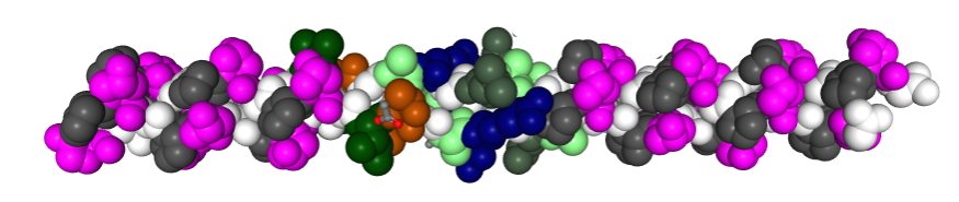
In order to make the correct number of bends in the collagen-like portion of the SP-D molecule diagram which I will use to overlay onto a TEM of one of the many published images of SP-D, i found that correspond to the glycine residues (in this image from RCSB PDB link that has a portion of a collagen molecule that can be viewed in 3D), and the comments on PDB-101, can be used as markers for the angular notations in said diagram.
This is a relief, and i compared the G– sequences in full length collagen (of which there is an easy to count number of at least 360 (that i counted by hand) and a very small number by comparison (29) in the SP-D portion. THus, i will put 29 bands in the diagram of SP-D which you will see is way less than the diagram of Girth Sorensen which I posted yesterday. which indicates there are 171 amino acids in the collagen-like domain. This is problematic, but here is the model of a collagen type triple helix I will use for the diagram.
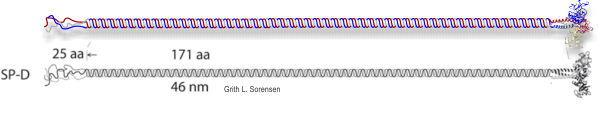
Redraw of the molecule SP-D from Sorensen
The best diagram i have found so far for surfactant protein D is by Girth Sorensen, and from his diagram I made one in color. I was not successful in finding any molecular structure on the protein database for the N terminal, and have many times sought help finding out about the collagen like domain. His diagram is pretty regimented, which is not likely to be accurate either because of the arc type shape seen in so many electron micrographs of those arms. It will try to find out how many repeats there are in the collagen like domain and create a wound structure that more approximates that number. In the meantime, it is almost laughable how many really bad diagrams of SP-D and SP-A there are in the literature…. the scientific literature, no less, some are really not informative. But i thought Sorensen’s was close.
One problem with this diagram is its rigidity. Other diagrams have this rigidity of the collagen-like domain as well, but add a quirk in the neck region, bending it at about a 45 degree angle – such a bend apparently located at the beginning of the coiled coil region. Since I have not found a single diagram with any molecular ribbon or ball and stick diagrams past the neck region, the depiction of a bend at that juncture is a little like fake news.
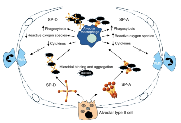
Truly embarrassing figure of the alveolar space
I belly laughed when i saw this diagram. It is supposed to represent an alveolus from a mammalian lung. Those long line things with circles, are they type I alveolar cells? ha ha… horrible perspective, and way out of proportion for the nucleus, and their thickness is too fat for the size of that pink thing at the bottom which they labeled alveolar type II cell. The nucleus of the type II cell looks to be drawn by a youngster, and the spirals (cut and carelessly pasted four times without any clue as to what they do or where they exit the cell — i will give them credit for at least putting them in the apical portion of the cell — which is still only half correct – LOL).
Just as cute is the SP-A diagram which in this case apparently doesn’t want the neck and collagenous regions to be “twisted” together as they are repeatedly reported (even by these same authors). SP-A and SP-D both look like the three portions of the CRD are one “ball”, which is inaccurate, even for the date of this publication…. that information was available. I wont complain about the depiction of the single blue alveolar macrophage, or the two polymorphonuclear leukocytes squeezing between type I cells but the black things in the alveolar space that look a lot more like millipedes or isopods or some other species of insect… than like bacteria or viruses— what!! that’s just not even funny, it is pathetic.
I am completely baffled why perfectly competent scientists can’t illustrate their findings with any degree of accuracy. I think they don’t even see how “off” they are.
\
Hilarious scientific diagrams
I have to laugh. There are few places in the visual world where copy-cat exists more than in scientific illustrations. Working on models of real and “artistic” diagrams of SP-D I can see sthis so clearly. Copy-cat as to mis-information as well as copy-cat as to color, and misnomers as labels and size of molecular elements. All of these diagrams are hysterical tributes to our willingness NOT to question truth. NOPE not talking politics here, but science.
Some of the clearly labeled SP-D diagrams look more like conglutinin diagrams.. than SP-D, and wouldn’t you know, when looking at shadowed conglutinin (Strang et al, Biochem J, 1986) they have a look more of SP-D with obtuse and acute angles. In fact the latter sometimes has an interesting configuration of three acute and two obtuse angles not always mirrored. SP-D seemed to be pretty regularly mirrored.
I am planning a comparison, that is, real measurements of the angular dimensions of SP-D from “TEM” images and angular dmensions of real-copy-cat measurementsof SP-D, to see how far adrift from reality we can go, even when we have reality (yes there is likely preparation and fixation artifact in the shadowed, negatively stained and AFM images- that is not new) right under our eyes.
Cover for CorelDRAW pro Dec 2008 snowflakes and hexagons
All these snowflakes used in a diagram (interlaced hexagons) were taken by a professor at Cal Tech. I contacted him for permission to use his “real” images. No monetary gain for me was derived, but I thoroughly enjoyed using the variety that nature has to offer and that he captured on camera and posted online. Also thanks to Mike Neer who is editor of this CorelDRAW help magazine, as he let me play around with images for a few of the covers.
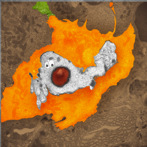
Click 5 – aka casper the friendly ghost
One fun image from long ago, never a cover illustration, but could have been one at some point, particularly for a fall issue of a throw-away-journal. This is actually from a micrograph taken from a KO mouse CLICK5. The particulars of that particular KO mouse have long passed from my memory, but constructing the image is still fresh. ha ha..
White part is the parietal cell nucleus, nose is red nucleolus) cytoplasm is pumpkin orange, and the green stem is a portion of of another cell.