Category Archives: Vesicles, isolates, fractions

Some pictures, not related to any project in particular that I can remember just some nice electron micrographs showing ultrastructure that hopefully will benefit someone

Weibel-Palade bodies: fun stuff

I have seen these… at the present moment I am looking over the published literature for Weibel Palade bodies (WPD) in order to determine if there is ever a place where ribosomes are present on the bounding membrane.  So far, none. This makes this cell adjesion group of molecules, while oligomerized into very beautiful structures, some like tubules, they are quite different from the intracellular granule purported to be a surfactant protein (possible SP-A) found in the cytoplasm of alveolar type II cells in the lungs of some species… which have a very precise localization and number of ribosomes on their limiting membrane (opposite sides of the long dimension of the granule, and approximately 4 ribosomes per 100nm).


Weibel-Palade Bodies (WPB) Layered granules in alveolar type II cells
At least 12 different proteins are found in granules Undetermined number of proteins in type II cell granules
Recovered as oligomers in clathrin coated vesicles Recovered, but not recycled or re-internalized as oligomers
Granules as single membrane bound SMOOTH ER Granules as single membrane bound with end to end distribution of ribosomes,
Interacts with cell elements in blood Interacts with fluid elements in alveolar space
Endothelial cell product Epithelial cell product
Interacts with white cells and platelets Interacts with alveolar macrophages
VWP apparently oligomerizes to influence the shame of WPBs Oligomerization of the SPs involved in intracisternal bodes of alveolar type II cells create the granuel shape
 WPBs maybe storage (albeit a small percent of total) for VWP Granules in type II cells maybe for surfactant protein storage
WPBs have P-selectin in small quantitites, i wonder if these are for binding of clathrin proteins to initiate recycling from the secretion bubble structures after exocytosis of VWP? Re-uptake, recycling of surfactant proteins is not so obvious (but happens) not big obvious vesicles…  at least not that I have seen.

Mouse, liquid breathing E2: macrophage and lysosomes

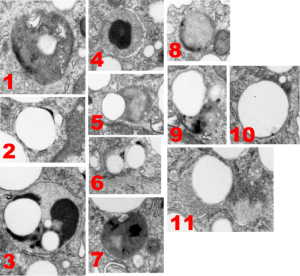
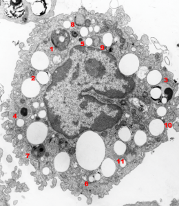
Here is a gallery of the types (big variation) of lysosomes that I found in a 1000A thick section of a single alveolar macrophage from a mouse that had breathed liquid E2 for 3 hours and then was allowed to recover for 17 days before euthanasia.

The only duplicate in style was found in the those MVB/LE/PFC (multivesicular body/late endosome/perfluorocarbon particle) that displayed a small very dense black cap, likely to be the distribution of the proteins.  Sometimes these contained one or two small droplets of E2 (6) or the densities were found at the margin of one large droplet of E2 (2) and even these have different morphologies. A less dense background and divided density contents of some MVB/LE/PFC bodies were NOT arranged with enzymes (proteins?) at the periphery only (3, 5, 7-9) and some polarity to the distribution of the darkest portion (more enzymes) of the MVB/LE/PFC. The lysosome 3 is quite interesting, with three definite PFC droplets and a very marked separation of a less electron dense background and dense compartment (presumably enzymes, maybe portions of some other proteins or even surfactant lipids.  Lipids would be a good guess since they are so darkly stained with the osmium tetroxide). The difference in the size of these droplets is interesting as well, since it seems that the smaller droplets (and maybe they shrink to not detectable with TEM) could be the ones which escape the cell and are offgassed in the lung.  I am just linking the complicated stuff on wikipedia about this, but you can be sure that the temperature difference between deep tissue and lung, and size differences in droplets makes a huge difference in vapor pressure…. someone else can figure this out).  Figure 5 below also shows the tiniest E2 droplets… actually interestingly also noted that the droplets are for the most part found on the periphery of the bounding cytoplasm (likely membrane bound, but perhaps the PFC just interfaces with adjacent proteins in a manner that looks membrane bound (that said for those droplets that do not LOOK to be inclusions in MVB/LE/PFC structures… but then, my guess is that they are all membrane bound having been taken in endocytically.

So the variation is huge, some, like image 1, look like there are lamellar body proteins resembling surfactant and lipids.

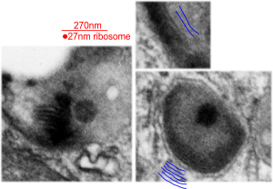
There are other MVB/LE/PFC structures which show a distinct lamellar nature with the distance from one dense line to the other is about about 32nm (see below). These are from 1, 9, and one not shown in the gallery above, but just cut off on the corner if image 1, but present in the whole alveolar macrophage at the bottom of this post.