Category Archives: Desmosomal mitochondrial associations

Mouse hepatocyte: mitochondrial desmosomal connections

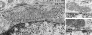
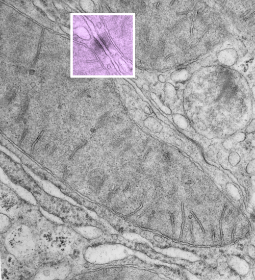
https://www.ncbi.nlm.nih.gov/pmc/articles/PMC2211412/pdf/nihms34115.pdfI have marveled at the mitochondria and their propensity to connect with both desmosomes and nuclear pore filaments. These little “bridges” in density and substructure connect them very often. Here is an hepatocyte sample just to show the point. Boxed area has mitochondria in one hepatocyte and mitochondrion in adjacent hepatocyte bridged by a desmosome on both sides.  There is great “order to the connection, first a lucent band (disk) then a dense and then a less dense, then a more dense and one more lucent band with little periodicities and a very dense and then lucent band and the plasmalemma.  So this is a total of 10-11 gradations in electron density  x 2, one on each side of the space between the hepatocytes, and within that space is of course the density pattern therin that is typical for desmosomes. The rigidity of the desmosomal “disk” or “round weld” is quite noticeable, on the plasmalemma side and the mitochondrial membrane side, and the thickness of the whole complex (from the membrane of the mitochondrion in one cell to the membrane in the adjacent cell is pretty fixed as well. I am pretty sure if you search PubMED you will find at least two publications where I mention this phenomenon. I don’t know why it has received so little attention.

This particular mitochondrion also has another connection with a desmosome about half a micron distal to this site.

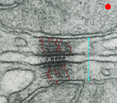
Making just a line diagram over top of the desmosome-mithcondrial connection i used just lines. Below that is a real great diagram of a desmosome with link to the publication. Ribosome=about 27nm depicted as a red dot. Blue arrow is about 200nm.s

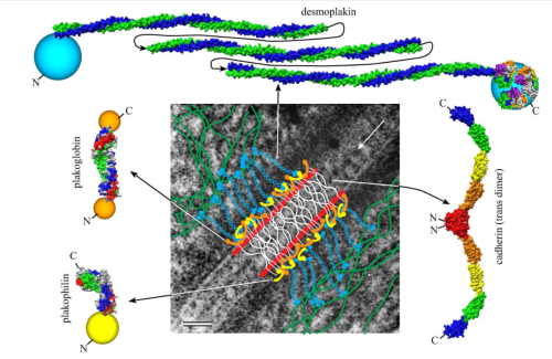
Manuscript was by DL Stokes.

Several examples of mitochondria – nuclear pore interactions

These nice interactions between nuclear pore outer filaments and the outer membrane of the mitochondrion have intrigued me for at least 3 decades.  They first came to my attention when looking at liver, hepatocytes specifically, where they are frequent, and uncluttered by a lot of other cell machinery and organelles.  It seems pretty obvious that their tethering an energy source to fuel translocation of cytoplasmic proteins into the nucleus: “proteins (such as DNA polymerase and lamins), carbohydrates, signaling molecules and lipids moving into the nucleus” according to wikipedia. The author of the wikipedia post suggests that 1000 translocations per pore complex occur each second. Smaller molecules diffuse through at the peripheral portions of the pore and specific nuclear localization sequences are recognized by the nucleoporin proteins to assist larger molecules through the central part of the pore channel. That author also suggests that the pore transport does not require energy, so there must be some function for the mitochondria found associated with nuclear pore complexes (and sometimes appear to be just bent and dragged toward them). It is possible that assembly and disassembly of nuclear pores would require mitochondria. It has been suggested that three NUPs which do occur on the outer (cytoplasmic) filaments get phosphorylated. It is likely a testable hypothesis, which could be ferreted out at least a little in examining different cell types and relating that to mitotic index, or alternatively, from unperturbed to apoptotic state.  It is not that unusual to find three different areas of the same mitochondrion to be seen associated with outer nuclear pore filaments.

One mitochondria (on the left) shows two such close encounters.  These examples come from either alveolar type II cells or hepatocytes; grey dots 27 nm ribosome, bar=270 nm. It is interesting that the nuclear basket (filaments on the nucleoplasmic side of the nuclear pore complex) are not visible in any of these pore-mitochondrial associations, except maybe slightly in image on lower right.  In each micrograph, the mitochondrion is top side, a portion of the nucleus is below.

Desmosomal-mitochondrial associations

The physical proximity of mitochondria to filaments in the cytoplasm has interested me for a long time. Particularly, those associations between mitochondria and the cytokeratin filaments (tonofilaments, intermediate filaments) on the cytoplasmic side of desmosomes and the associations between mitochondria and nuclear pore filaments (those projecting from the cytoplasmic face of the pore into the cytoplasm). Anchoring plaque proteins and central proteins are pretty nice in this electron micrograph which wikipedia describes thusly: “transmembrane linker proteins, called desmoglein and desmocollin, belong to the cadherin family of cell-adhesion molecules bind to plakoglobin and other proteins in the plaques and extend into the intercellular space, where they interact, forming an interlocking network that binds two cells together”.

I have hundreds of examples of these interactions, the former being more numerous in some cell types, the other more numerous in others. Here is an electron micrograph of one particularly nice association between a mitochondrion and a desmosome.  There can be mitochondria on both sides of a desmosome as sectioned in 2D, which begs the question of the 3D aspects of these associations.

electron micrograph of mitochondrion associated with desmosome

In fact, a single mitochondrion can be associated with more than one desmosome, and can also touch on the cytoplasmic-side-filaments of more than one nuclear pore.  M, mitochondrion, arrow, desmosome, bar= 100nm dot is 27 nm by ribosome. Mouse liver female control C4.