Category Archives: Perfluorodecyl iodide

Perfluorodecyl iodide: a peroxisome proliferator

Going through archived images of the livers of mice which received infusions of iodo containing perfluorochemicals, I ran onto a hand-written comment which made me laugh. It was “funky print given under pressure to LC Clark (Jr) that perfluorodecyl iodide increases tremendously the number and size of microbodies (aka peroxisomes) in the mouse liver”  (words in italics added today for clarity).  Apparently no one wanted to hear that news in 1973 or 4… perhaps because it was a “damning” mark on the possible use of  perfluorochemical formulations in general, and specifically for artificial – blood substitutes.

Wikipedia summary is here: “They are involved in catabolism of very long chain fatty acids, branched chain fatty acids, D-amino acids, and polyamines, reduction of reactive oxygen species – specifically hydrogen peroxide; – and biosynthesis of plasmalogens, such as ether phospholipids critical for the normal function of mammalian brains and lungs. They also contain approximately 10% of the total activity of two enzymes in the pentose phosphate pathway, which is important for energy metabolism.”
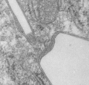
Below are three images, one of liver which shows the very large peroxisomes, in this particular micrograph (not all shown here) they were a volume density of about 30% of the cytoplasm which puts IPFD up there with the best of the proliferators; mitochondria for comparison were about 33% of the cytoplasm. Consensus has peroxisomes at more like 2% of the volume density of the hepatocyte under unstimulated conditions.

The story will be complicated as there are more than 30 enzymes which are in peroxisomes: and the patterning that is seen in the enzymes within the phagolysosomes containing IPFD looks to be similarly wound as the enzymes within peroxisomes in the hepatocytes (top two image groups). From the internet the middle three images (used without permission as a screen print of the insanely egotistical Dr. Jastro TEM collection who most certainly used public funds for this research and collection of micrographs and yet thinks he has ownership of these images…. not civil at all in my opinion and shame on him) is an hepatocyte peroxisome (which i measured against a nearby ribosome and also the micron bar marker, for size) shows a coarse texture, somewhat coiled. The latter and my peroxisome images (top), added from a portion of an hepatocyte from the liver which received IPFD and measured against a nearby ribosome, show similar “oligomerization” or “substructural” protein patterning.

Some protein(s) is creating this texture. It would be interesting to know if the protein in these microbodies (aka peroxisomes) is the same as that which is found in abundance in the phagolysosomes containing IPFD. Bottom micrograph is a portion of hepatocyte cytoplasm from IPFD infused mouse more than 200 days post infusion from which my (top two) images are derived.
electron micrograph hepatocyte peroxisome proliverator perfluorodecyl iodide

Finding the crystal width pattern for perfluorodecyl iodide

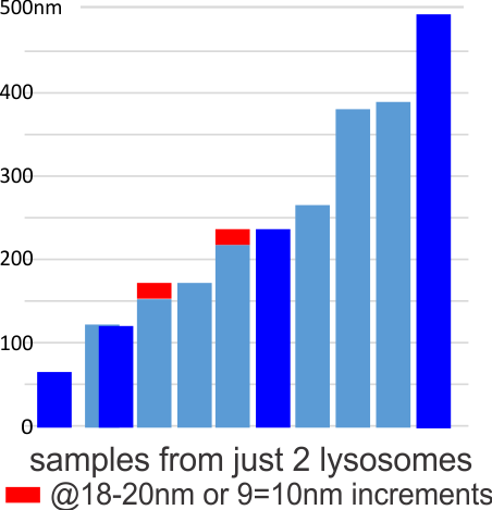
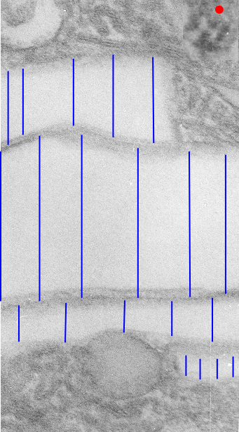
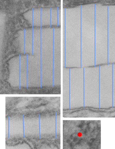
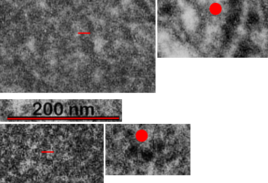
I have felt there was a pattern to the width of the perfluorodecyl iodide crystals in the micrographs that I have for some time. Today I decided to measure just two sets of crystals from two different lysosomal structures from two different cells. The graph below shows that there might be a stepwise increase to the thickness of the crystals of something on the order of 18-20nm (or maybe half that). The data from 4-8 measurements along the wide-axis of individual crystals were measured and normalized to the 27nm size of an adjacent ribosome. The mean widths sorted in ascending array and the second plot measured identically (had a few more crystals to measure) is superimposed. The red rectangles might indicate an incremental increase in size. Just thinking on this. I did find a couple of publications on “incremental pattern mining” which would be fun to examine.  Hopefully I will find someone with software already in place to do this. The actual micrographs and positions of measurement are shown below.  Red dots=27nm a presumed size for a mammalian ribosome, in each micrograph. Dont miss the lysosomal protein “snake-like protein with dark central stripe” mentioned in previous which is very clear in the top portion of the top electron micrograph. In this micrograph the values are 18-20 and 9-10 nm respectively for the two-band protein (similarly to previous post HERE).

Iodoperfluorocarbons

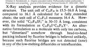
I found a very old article, portion below I did not retype but added the reference at the top, which mentioned briefly what I think is perfluorodecyl iodide (1-iodoperfluorodecane). I read this to mean that it is crystalline in nature and that the molecule has dimensions which might indicated that it has dimerized.  If I knew what a “unit cell” was I might be able to relate it to the size (which is given here in Angstrom units) of 15.5 to 16,  I might be able to figure out whether the periodicity found at the long ends of the IPFD crystals in vivo  (within lysosomal structures) and the round organization seen in some images have relevance to that dimension.

I found a more current article which seems to rehash some of what this early article by Rondesvedt mentioned in 1969. I like that he mentions that iodoperfluoroalkanes are bio-persistent, and that fits with the “mouse emulsion infusion” data that I see.  That second publication is here.  But there is no way without learning chemistry I can figure it out.

And in terms of environmental impact of PFC, in particular PFOS and PFOA and other perfluorochemicals in wide-spread usage, this is a nice article to read.

from which is a nice quote “Since the first reports revealing the widespread global occurrence of PFOS in wildlife (Giesy and Kannan 2001) and the frequent detection of PFASs in human blood (Hansen et al. 2001) were published a decade ago, the scientific literature on the environmental and toxicological aspects of PFASs has burgeoned rapidly, and the rate of publication currently exceeds 400 articles per year. In the existing body of literature, including governmental reports, authors have created terminology, names, and acronyms to describe these substances. Unfortunately, inconsistencies have inevitably arisen between various groups of authors. In the absence of any concerted effort between scientists to agree on a common terminology to designate the substances, a given compound has often been denoted by a variety of different names and acronyms, or a given acronym has been used to represent different substances. In addition, names to describe broad groups of substances have proliferated that in some instances mistakenly portray substances that are very different from one another as being the same. As a result, the scientific literature for these substances has at times become confusing. There is a need for harmonized terminology, names, and acronyms that clearly and specifically describe PFASs.” showing how difficult it is to read the literature on these chemicals.

I had to look up the words “unit cell” not being a chemist I had no clue what that meant… so here is a definition — A unit cell is the most basic and least volume consuming repeating structure of any solid. It is used to visually simplify the crystalline patterns solids arrange themselves in. When the unit cell repeats itself, the network is called a lattice.

Substructure of the lysosomes containing perfluorodecyl iodide

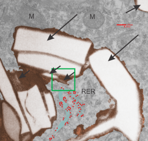
Substructure of the lysosomes containing perfluorodecyl iodide is very interesting. There are issues both with the lysosomal membrane, which in some places may be continuous with other membranes, in particular, i think maybe the RER. Because perfluorocemicals are kind of “slippery” and I think they can travel easily retrograde-style back up into the RER, then it makes sense that some profiles of RER would be seen at sites within the lysosome periphery and the adjacent cytoplasm. I haven’t been able to equivocally find ribosomes on the actual lysosomal membrane to verify that concept.  There are perfluorodecyl iodide tiny crystals…. this is interesting…. an obvious type of fracture or separation plane along the long axis of the larger crystal inclusion certainly is evident….  and i am more convinced that the pattern of lysosomes (a dotted or tubular pattern) is on the order of 20-30nm diameter or width.

Here is an electron micrograph: mouse, neg 9722_10%IPFD, 5%F68 infused 100cc/kg,   8-14-73 sac 5-8-74, 267 days post infusion;  liver. Brown areas=lysosomal enzymes within a bounding membrane, white=IPFD, red dots are ribosomes; pale green is cisterna of RER, green box is area enlarged below.

perfluorodecyl iodide transmission electron micrograph lysosomes

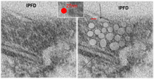
So the bottom two images, left unretouched, right unretouched but with translucent overlays where I see a pattern within the lysosomal enzyme membrane.  Arrows here point to fractured off crystals of IPFD, and the red line is a marker for 27nm which is the same size as the ribosome measured (top inset)…. this makes me wonder if there is some connection between this pattern and lysosomal contents.

Periodicity at ends of IPFD crystals in macrophages

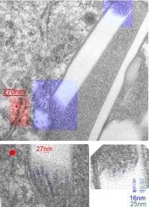
I looks like the lysosomal enzymes at the end of the IPFD crystal inclusion-structures might be tubular. But then as the enzymes pass away from the IPFD itself, there can be a coiling up of the protein with less rigidity.  Measurements at ends of IPFD look smaller than those in the lysosom proper… may be error of just having only measured a single crystal. Same negative and block as previous post, and the ends of the IPFD crystal are colored bluish, and the area measure for ribosome size is in red. Enlargements below, with areas specific measured highlighted. The dense part of the period and the lucent areas beside it.

Texture comparisons: background, cytoplasm and IPFD-lysosome

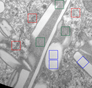
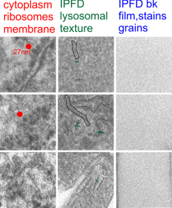
I am trying to determine whether there is a predictable texture to the proteins within phagolysosomes which contain IPFD (perfluorodecyl iodide)(which I am positive there is–so this is academic). I have compared territories from one micrograph with marked inset boxes: red boxes=cytoplasm with a ribosome size marker for comparisons of texture-size); green boxes=portions of the compact and textured lysosomal proteins found just within the limiting membrane of the phagolysosomes which contain IPFD; blue boxes=the background texture of the micrograph found over the “empty” IPFD granules which consists of the acetate film grain and the stains– osmium, uranyl acetate and lead citrate grains. No photographic paper grains are present as this was scanned at 5000 ppi from the negative.

I measured a similar texture pattern from the end-on portion of the lysosomal proteins in last post, and dimensions are similar but not identical… likely my fault, not the fault of the proteins.  So todays measurements are something around the following: dense areas at around 17nm (corresponding to the “spot” in yesterdays post which was around 13nm) and the surrounding more electron lucent space measures about 34nm (which in yesterdays post was closer to 40nm)–all likely resulting from variations in how i measured ribosome size, which is the benchmark.

Image below is from a randomly selected phagocyte in the liver (I cannot identify whether this is a Kupffer cell or other phagocytic cell type). These cells can be closely aligned with sinusoids but also near portal tetrads. They are typically “overflowing” with IPFD particles. IPFD stays around for months, unlike other some perfluorochemicals.

Neg 9722 block 3775 10% IPFD 5%F68 injected 8 14 1973 100cc/kg sac 5 8 1974 mouse liver 267 days.

perfluorodecyl iodide electron microscopy 267 days post infusiontexture of lysosomes containing perfluorochemical crystals of perfluorodecyl iodide

IPFD crystals in lysosomes

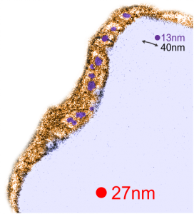
RedI cut around the top end of the crystal inclusion in the image from the previous post, and erased (in photoshop) areas that allowed me to pseudocolor the objects that I could see in the lysosomal phase of this phagososome. I think the periodicity is going to be interesting with a 13-15nm round central dense area and a lucent area around that density (like a donut), still with a layer of dense protein or a trilaminar membrane.

It was fairly easy to see the repeating dots and lucent surroundings emerge as a repeating pattern along the terminal membrane at the long axis of the IPFD inclusion. IPFD crystals show for the most part a definite polarity, and any substructure in the IPFD inclusion itself is see on the butt ends of the short width axis, while a very smooth bounding area is seen on both sides running in a parallel direction.

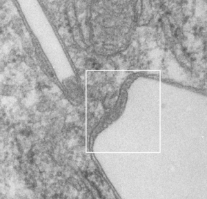
Below, same pix as 9 13 2017 post but with a bounding box which aligns to the inset image below again.


Red dot=ribosome at 27nm diameter, purple dot=central densities, tangential end sections, 40nm dimension for a cross sectional diameter of the electron lucent area surrounding the purple dot, and 13 nm dimension is for the purple dots (some larger some smaller).

Awesome IPFD crystals in lysosomes

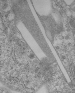
Perfluorodecyl iodide was examined once upon a long time ago as a possible PFC for blood substitutes. There are a couple of tissue blocks which I am re-examining to see whether there is a periodicity to the lysosomes that are included in these amazing phago-lysosomal structures, left over footprints as it were, to the presence of IPFD in macrophages (Kupffer cells, fat storing cells?, circulating macrophages, in the liver).

perfluorodecyl iodide transmission electron microscopyperfluorodecyl iodide transmission electron microscopy
This lower micrograph has very definite structure at the ends (one top one bottom) of the crystals. It is a little unfortunate that at the time i printed these negatives (back in the ‘wet darkroom days’ that I didnt use a finer grade paper and developer. Too much grain, but the little indents and rounded areas with a punctate density are going to be relly interesting. I think the lucent areas beside each will measure out the same as some fine lines that I see at the ends of other crystals.