Monthly Archives: October 2018

Quintessential SP-D dodecamer image

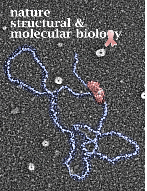
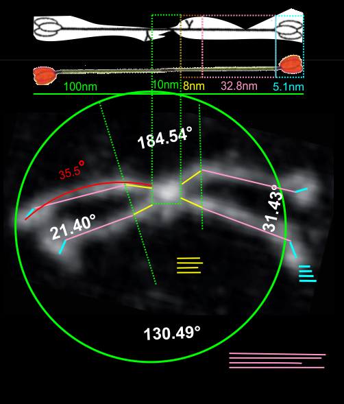
Measuring 30 SP-D dodecamers derived from numerous publications using several different methods for visualization, led me to identify this particular image as the quintessential SP-D dodecamer. The measurements then, from this particular image (most likely from the many images produced by Crouch et al, but which I have also upped the ppi and contrast on) had the closest arc’s angles in each of the individual trimer arms comprising the dodecamer to that which I measured from around 120 trimers. That is…right around 35 degrees.  The distance to what looks like a bend and a bright spot in each of the trimeric arms in the electron micrograph likely represent the glycosylation site which was identified by several different investigators.  This point gives the trimers the look of an arc. The carbohydrate recognition domains as they attach to the “neck” also like an angle (not yet measured). The center “bright area” of the dodecamer area where all the trimers are bound at the N terminal has a width of about 10nm, at least as is measured in this quintessential image (A collective measure the width and length of the central core of the SP-D dodecamer needs to be done). The the mean for the acute and obtuse angles of the four arms (about 30-50o and 120-140o — in this case the angles are 21.4  and 31.43 and the obtuse angles are 184.54 and 130.49 and so are a not exactly representative but close). 99% confidence level is between 37.00 and – 58.39o for the two acute center angles.

Two commonly reproduced diagrams are included at the top of the real SP-D for comparison. It is obvious that the flexibility of the SP-D dodecamer is clearly missed. And in support of the arms of the dodecamer NOT being stiff, but flexible, authors have suggested that mutant SP-D molecules (shortened collagen-like domains, deleted collagen-like domains, etc) can retain some functions in innate immunity, though sometimes differently than the native SP-D.

quintessential surfactant protein D dodecamer image

The purpose for making the measurements is to determine whether the fuzzyball structures made of dodecamers joined in the center at have the straight arms as depicted in nearly every single SP-D diagram.
Arc angle of trimeric arm(s) of CL-43 compared to SP-D

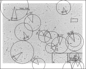
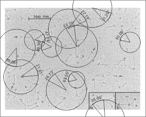
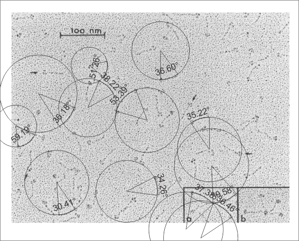
This is a quick test of the arc angle of the trimeric arms of three different collectins but mostly CL-43. The measures are made from publication(s) from other investigators but for this post, principally from  Holmskov et al, 1995 so these are not my micrographs. Each molecule (individually) for CL-43 and bovine conglutinin and SP-D seem to display  slightly curved trimeric arm(s) (totally unlike the diagrams of SP-D, conglutinin and CL-43. These diagrams are typically straight lines arranged as X s clearly not right. This discrepancy was something that grabbed my interest).  Quickly measured arc angle of CL-43 molecules  (n= 35) shows an arc angle of  38.3+/1 1.79. This was not significantly different than a couple of dodecamers of SP-D measured similarly (using the best fit circle and arc length to determine the angle) which was  n=11 molecules,  x=35.7+/- 1.71.  I will add the arc angle of conglutinin molecules and a couple of measures for mutant SP-D from papers published in the scientific literature. But here is the single image (replicated 3 times for different measures of the arc angle of CL-43 as shown in the black lines and degree text in each image.  The insets are SP-D and conglutinin, with just one molecule each in that inset. Molecules were rotary shadowed according to the authors M&M.

A paired dataset, now with many more samples of SP-D angles than CL-43 shows the two very nearly the same.

currently: ONLINE calculator gives these values
Surfactant protein-D
(19 dodecamers – 4 arms measured independently)
N1: 75
df1 = N – 1 = 75 – 1 = 74
M1: 38.11
SS1: 10426.67
s21 = SS1/(N – 1) = 10426.67/(75-1) = 140.9
CL-43
(36 measures = 6 dodecamers – 4 arms measured independently)
N2: 35
df2 = N – 1 = 35 – 1 = 34
M2: 38.32
SS2: 3847.39
s22 = SS2/(N – 1) = 3847.39/(35-1) = 113.16

The results are show no significant difference in the arc angle in the collagen portion between these two molecule types. The next thing would be to compare the aa sequences and aa length of the collagen domain of the arms of these two different collectins. In fact with the current dataset…. the SP-D is right on 38.1 degrees just like CL-43. The calculations us each of the four angles per dodecamer as an N so n= “arc angle”

THE FINAL t-test are as follows:

N1: 35
df1 = N – 1 = 35 – 1 = 34
M1: 38.32
SS1: 3847.39
s21 = SS1/(N – 1) = 3847.39/(35-1) = 113.16

Treatment 2

N2: 129
df2 = N – 1 = 129 – 1 = 128
M2: 35.72
SS2: 20437.31
s22 = SS2/(N – 1) = 20437.31/(129-1) = 159.67
WHETHER calculated with the n of SP-D molecules individually (that is 4 per dodecamer – yes there was one molecule where i could only measure the arc angle of three arms) or whether the t test was done using the mean of the arc angles for the dodecamer as N, where n=30 for SP-D, the results were almost identical.

it will be interesting also to see whether conglutinin is similar…

What to measure, what to compare

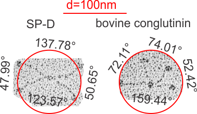
In looking at the morphology of a few of the innate immune proteins (collectins) I have been trying to sort out whether the collagen-like domains have a “similar” flexibility, or tendency to “bend”.  I was hoping to find some differences, but if they exist I think they will be small (though small does not preclude significance). I found one publication that showed rotary shadowed molecules of CL-43, or SP-D and bovine conglutinin… hopefully  at equivalent magnifications (as stated). The micrograph showed trimers of CL-42, dodecamers of SP-D and conglutinin.  There is a slight curvature to the collagen-like domain (which is invariably NOT shown in diagrams) which is pretty consistent in micrographs from many publications.

The purpose is to determine if the slight curvature has bearing on how the SP-D multimers associate, and how their trimeric arms are positioned in “fuzzyballs”.  The arc angles are shown for each quadrant of SP-D and conglutinin below. THe CL-43 is not shown because it apparently does not form dodecamers.

I am also linking a site for calculating arc angles and other measures of circles as a really helpful teaching website.

Hands IN Food BIN

Does anyone but me see a problem here. Better question, does anyone anymore recognize the germ theory of disease to be valid. Or is everyone a “natural” scientist and have their own opinion.
Kroger has put out refrigerated bins (i think to copy Whole Foods) but do you see that the tongs that are used to individually pick up each item are just put back on top of the FOOD itself. Dirty hands, hands that have just picked noses, wiped babys drool, put their fingers around dirty Kroger shopping carts, handled raw meat packages. WOA… something is not right here. Wake up Kroger.

Creating an accurate diagram of SP-D —

In order to make the correct number of bends in the collagen-like portion of the SP-D molecule diagram which I will use to overlay onto a TEM of one of the many published images of SP-D, i found that correspond to the glycine residues (in this image from RCSB PDB link that has a portion of a collagen molecule that can be viewed in 3D), and the comments on PDB-101,  can be used as markers for the angular notations in said diagram.

This is a relief, and i compared the G– sequences in full length collagen (of which there is an easy to count number of at least 360 (that i counted by hand) and a very small number by comparison (29) in the SP-D portion.  THus, i will put 29 bands in the diagram of SP-D which you will see is way less than the diagram of Girth Sorensen which I posted yesterday. which indicates there are 171 amino acids in the collagen-like domain. This is problematic, but here is the model of a collagen type triple helix I will use for the diagram.


Redraw of the molecule SP-D from Sorensen

The best diagram i have found so far for surfactant protein D is by Girth Sorensen, and from his diagram I made one in color. I was not successful in finding any molecular structure on the protein database for the N terminal, and have many times sought help finding out about the collagen like domain.  His diagram is pretty regimented, which is not likely to be accurate either because of the arc type shape seen in so many electron micrographs of those arms.  It will try to find out how many repeats there are in the collagen like domain and create a wound structure that more approximates that number.  In the meantime, it is almost laughable how many really bad diagrams of SP-D and SP-A there are in the literature…. the scientific literature, no less, some are really not informative.  But i thought Sorensen’s was close.

One problem with this diagram is its rigidity. Other diagrams have this rigidity of the collagen-like domain as well, but add a quirk in the neck region, bending it at about a 45 degree angle – such a bend apparently located at the beginning of the coiled coil region.  Since I have not found a single diagram with any molecular ribbon or ball and stick diagrams past the neck region, the depiction of a bend at that juncture is a little like fake news.

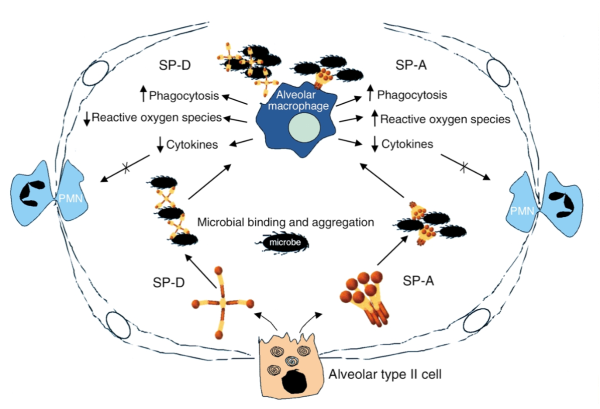
Truly embarrassing figure of the alveolar space

I belly laughed when i saw this diagram. It is supposed to represent an alveolus from a mammalian lung. Those long line things with circles, are they type I alveolar cells?  ha ha… horrible perspective, and way out of proportion for the nucleus, and their thickness is too fat for the size of that pink thing at the bottom which they labeled alveolar type II cell. The nucleus of the type II cell looks to be drawn by a youngster, and the spirals (cut and carelessly pasted four times without any clue as to what they do or where they exit the cell  — i will give them credit for at least putting them in the apical portion of the cell — which is still only half correct – LOL).

Just as cute is the SP-A diagram which in this case apparently doesn’t want the neck and collagenous regions to be “twisted” together as they are repeatedly reported (even by these same authors).  SP-A and SP-D both look like the three portions of the CRD are one “ball”, which is inaccurate, even for the date of this publication…. that information was available.  I wont complain about the depiction of the  single blue alveolar macrophage, or the two polymorphonuclear leukocytes squeezing between type I cells but the black things in the alveolar space that look a lot more like millipedes or isopods or some other species of insect… than like bacteria or viruses— what!!  that’s just not even funny, it is pathetic.

I am completely baffled why perfectly competent scientists can’t illustrate their findings with any degree of accuracy.  I think they don’t even see how “off” they are.

\

Diagrams of SP-D dodecamers vs the real thing

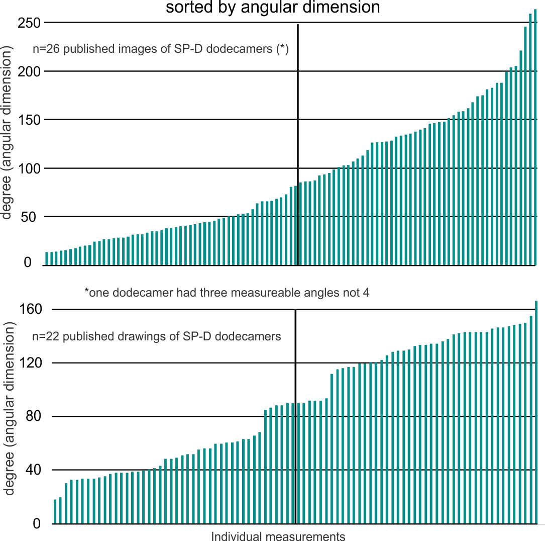
Trying to determine where there is any rhyme or reason for the angles that individuals choose when they diagram SP-D dodecamers I decided to compare some actual measurements vs some drawings. Each and all images come from published (peer reviewed) articles available to anyone who searches the internet for surfactant protein D.

Briefly, there seems to be a huge variation in angular dimension in the photos taken using various types of electron microscopy (including atomic force microscopy), more variability in fact than the diagrams would represent.  Initially, the diagrams that represent SP-D as an X with 4 90o angles are not the most accurate, as the “mode” for angles from measuring “real” images is about 30-50o and 120-140o. Because of the variability in angle dimensions (and a publication by Arroyo et al demonstrated something along these lines using pH as a variable to alter the angular dimension of SP-D dodecamers (and multimers)) one has to think about environmental factors in the shape of this protein. In the search for a good pattern recognition protein to attach to nanospheres for immunity… the hunt for the optimal angle(s) is important.

Below is the set of measured angular dimensions for SP-D. A summary sheet for the “real” SP-D images from several different authors (including, Waters et al, Crouch et al, Hildago et al, Hseih et al, Ohya et al, Kishore et al,  Arroyo et al, to name a few) follows.  It becomes clear that the division of SP-D dodecamer arms into acute and obtuse angles as portrayed by the authors in their diagrams may not replicated in the real life continuum, likely due to environmental influence (including pH as described by Arroyo et al, but other factors as well).

Blood glucose levels and lichen planus

I just found this pdf, (attached below) and in my own experience with lichen planus have seen some carbohydrate metabolism issues, though mainly I get more hypoglycemic (self diagnosed, it might be hyperglycemia) than I have in previous years. Also, just the level of fatigue is increased (but also, i am 74 and expect to be able to easily run marathons)..Keeping all in perspective, i am just trying to get to the best way to live with this diagnosis. IJCED_3(1)_14-16> find pdf here
Five years down the road (it is now May 2023): This has never gone away, so i guess i am on the long end of this disease (commented at 7 yr in the literature) than the short end (1 year) and for me “stress” is certainly a factor, and there is a possibility that working with concrete and mortar might make things worse? No reasons for that thought except coincidence? since I have spent the winter and early spring patching basement holes, and tiling a bathroom. Who could make a connection? no clue.

Injury to superficial skin (like scraping injuries that remove epidermis) are definitly a contributing factor.